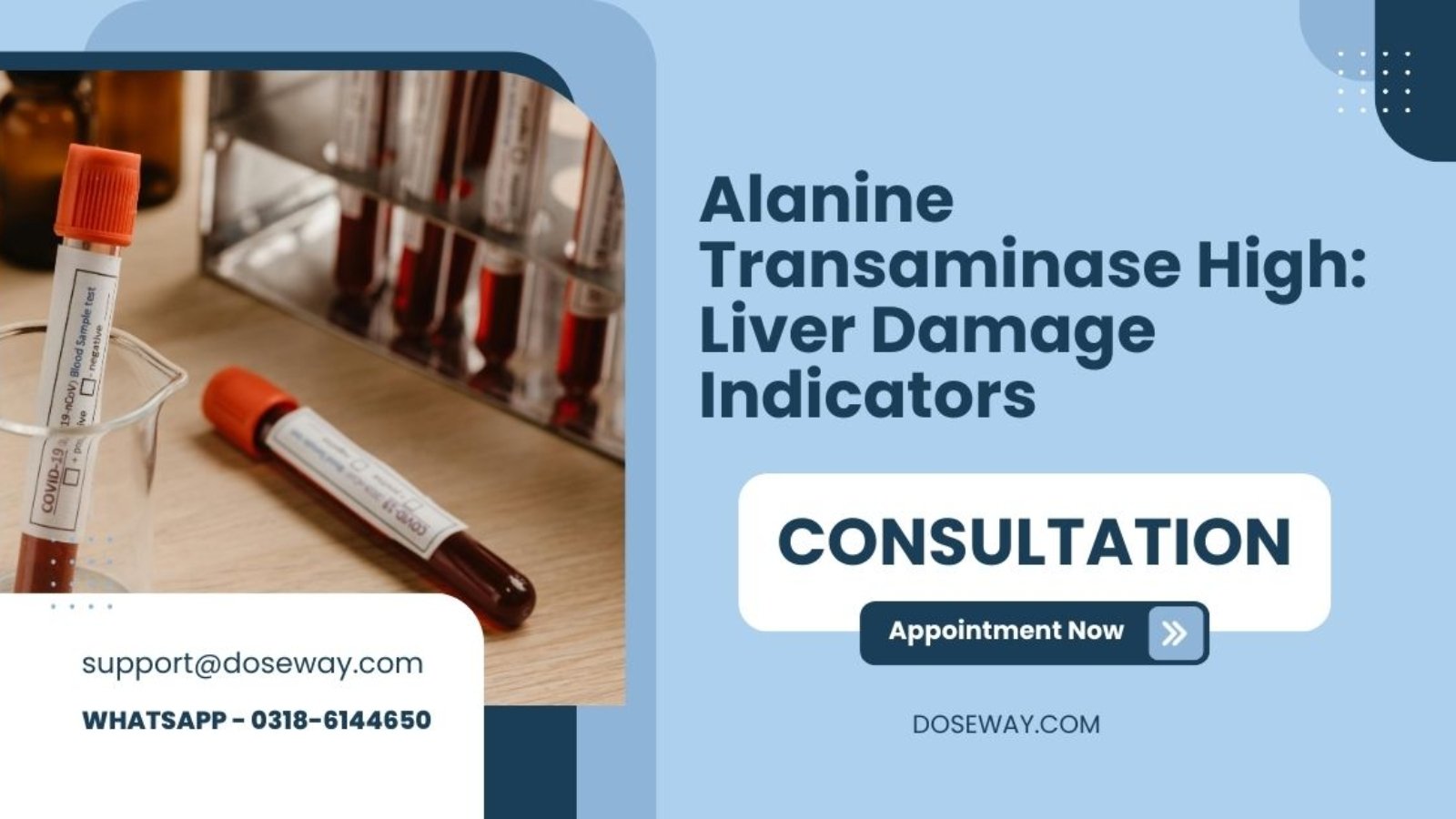
Alanine-Transaminase-High-Liver-Damage-Indicators

Alanine Transaminase High: Liver Damage Indicators

Alanine transaminase (ALT) is a crucial liver enzyme that serves as a primary indicator of liver health and function. When liver cells become damaged or inflamed, they release ALT into the bloodstream, making elevated ALT levels one of the most significant markers of potential liver damage. This comprehensive guide explores everything you need to know about ALT levels, liver function indicators, and how to interpret your liver health metrics using our advanced Liver Function Calculator.

In this definitive guide, you’ll discover:

• The critical role ALT plays in liver health assessment
• How to interpret your liver function test results accurately
• Common and rare causes of elevated ALT levels
• Practical strategies to improve your liver health naturally
• When to seek medical attention for abnormal liver readings
• How to use our free liver function calculator for personalized insights

ALT Levels & Liver Function Estimator

ALT Levels & Liver Function Estimator

1
Demographics
2
Medical History
3
Liver Tests
4
Results

Personal Information

Medical History & Lifestyle

/

Liver Function Test Results

0 U/L
0 U/L
0 U/L
0 mg/dL
0 g/dL
0 U/L

Liver Function Assessment

Normal
0
Score

Interpretation

Based on your inputs, your liver function appears to be within normal ranges. Continue maintaining a healthy lifestyle.

Liver Health Indicators

ALT Level 0 U/L
AST Level 0 U/L
ALP Level 0 U/L
Bilirubin Level 0 mg/dL

Disclaimer: This tool provides estimates based on standard medical guidelines and should not replace professional medical advice. Consult a healthcare provider for accurate diagnosis and treatment.

Read More Guide & Try Free Tools:

Free-Alanine-Transaminase-High-Liver-Damage-Indicators

What is Alanine Transaminase (ALT) and Why Does It Matter for Liver Health?

Alanine transaminase, commonly known as ALT, is a vital enzyme predominantly found in liver cells that plays an essential role in amino acid metabolism and energy production. When liver cells experience damage or inflammation, ALT leaks into the bloodstream, making elevated levels a primary indicator of hepatocellular injury. Medical professionals consider ALT one of the most liver-specific enzymes, with levels that directly correlate with the degree of liver cell damage. Understanding your ALT levels provides crucial insights into your liver’s functional status and overall metabolic health.

The significance of ALT testing extends beyond simple liver screening. This enzyme serves as an early warning system for various hepatic conditions, from mild fatty liver to more severe conditions like hepatitis or cirrhosis. Regular monitoring of ALT levels enables healthcare providers to detect liver abnormalities before symptoms become apparent, allowing for early intervention and prevention of progressive liver damage. At Doseway, we emphasize the importance of understanding these fundamental liver markers through our comprehensive online doctor consultation services.

Key facts about ALT every person should know:

• ALT is primarily concentrated in liver cells, with smaller amounts in kidneys and heart
• Normal ALT levels typically range between 7-56 units per liter (U/L)
• Men generally have slightly higher normal ALT ranges than women
• ALT levels can fluctuate due to medications, alcohol consumption, and dietary factors
• Persistently elevated ALT requires medical investigation regardless of symptoms
• Our lab tests at home service makes ALT monitoring convenient and accessible

How Does the Liver Function Calculator Work and What Parameters Does It Assess?

Our advanced Liver Function Calculator represents a sophisticated digital health tool designed to evaluate multiple aspects of liver health through a comprehensive assessment of clinical parameters, lifestyle factors, and biochemical markers. This innovative calculator processes your specific health data through validated medical algorithms to generate personalized liver health risk assessments and actionable recommendations. By analyzing both laboratory values and modifiable risk factors, the calculator provides a holistic view of your hepatic wellness that empowers informed health decisions.

The calculator assesses three primary categories of health information: demographic characteristics, medical history and lifestyle factors, and specific liver enzyme values. Each parameter carries weighted importance in the final risk calculation, with ALT levels typically receiving the highest consideration due to their liver-specific nature. The algorithm processes these inputs against established medical reference ranges and clinical guidelines to determine your individual liver health status and potential risk levels. Through Doseway‘s digital health platform, you can access this sophisticated assessment tool alongside our complete suite of health services at your doorstep.

The calculator evaluates these critical liver health parameters:

• ALT (Alanine Aminotransferase) levels and their deviation from normal ranges
• AST (Aspartate Aminotransferase) values and the AST/ALT ratio significance
• ALP (Alkaline Phosphatase) measurements for bile duct health assessment
• Bilirubin levels indicating liver processing and excretion efficiency
• Albumin production as a marker of liver synthetic function
• GGT (Gamma-Glutamyl Transferase) for alcohol-related liver impact
• Lifestyle factors including alcohol consumption, medication use, and dietary patterns

What Constitutes Normal ALT Levels and When Should You Be Concerned?

Normal ALT levels demonstrate significant variation based on multiple factors including age, gender, body composition, and laboratory reference standards. Generally accepted normal ranges fall between 7-56 units per liter, with women typically showing lower baseline values than men. Recent research suggests that optimal ALT levels might be even lower than traditionally accepted ranges, with values under 30 U/L for men and 19 U/L for women representing truly healthy liver function. Understanding these nuances helps interpret your liver enzyme results within the appropriate clinical context for your specific demographic profile.

Concern regarding ALT levels escalates when values exceed the upper normal limit, particularly when elevations persist on repeated testing. Mild elevations (1.5-2 times upper normal limit) often warrant monitoring and lifestyle modification, while moderate elevations (2-5 times normal) typically require medical investigation. Severe elevations (5-10 times normal or higher) represent medical emergencies requiring immediate attention. Through Doseway‘s accessible online pharmacy and telehealth services, individuals with abnormal ALT levels can conveniently access professional guidance and necessary medications to support liver health.

ALT level interpretation guidelines:

• Optimal range: Below 30 U/L for men, below 19 U/L for women
• Mild elevation: 1.5-2 times upper normal limit (approximately 56-112 U/L)
• Moderate elevation: 2-5 times upper normal limit (approximately 113-280 U/L)
• Severe elevation: 5-10 times upper normal limit (approximately 281-560 U/L)
• Critical elevation: Over 10 times upper normal limit (above 560 U/L)
• Persistently elevated levels require investigation regardless of degree

What Are the Most Common Causes of Elevated ALT Levels?

Elevated ALT levels originate from diverse etiologies ranging from temporary, reversible conditions to chronic, progressive liver diseases. Non-alcoholic fatty liver disease (NAFLD) represents the most common cause of mildly to moderately elevated ALT in developed countries, affecting approximately 25% of the global population. This condition involves excessive fat accumulation in liver cells, triggering inflammation and cellular damage that releases ALT into the bloodstream. Other prevalent causes include alcohol-associated liver disease, medication-induced liver injury, and viral hepatitis infections, each requiring distinct diagnostic approaches and management strategies.

Less common but clinically significant causes of ALT elevation include autoimmune hepatitis, Wilson’s disease, alpha-1 antitrypsin deficiency, and celiac disease. Even non-hepatic conditions like muscle injury, thyroid disorders, and strenuous exercise can transiently elevate ALT levels, highlighting the importance of comprehensive clinical evaluation beyond isolated laboratory findings. At Doseway, our integrated healthcare approach through online doctor consultation services ensures proper identification of ALT elevation causes and development of personalized management plans.

Primary causes of elevated ALT levels:

• Non-alcoholic fatty liver disease (NAFLD) and its progressive form NASH
• Alcohol-related liver injury and alcoholic hepatitis
• Viral hepatitis (Hepatitis A, B, C, D, and E viruses)
• Medication-induced liver injury (acetaminophen, statins, certain antibiotics)
• Autoimmune liver conditions (autoimmune hepatitis, primary biliary cholangitis)
• Metabolic disorders (Wilson’s disease, hemochromatosis, alpha-1 antitrypsin deficiency)
• Celiac disease and other intestinal conditions affecting liver function

How Do You Interpret ALT Levels in Relation to Other Liver Enzymes?

ALT interpretation gains significant clinical meaning when analyzed in conjunction with other liver enzymes, particularly AST (aspartate aminotransferase). The AST/ALT ratio, known as the De Ritis ratio, provides valuable diagnostic clues about the underlying cause of liver injury. A ratio less than 1 typically suggests non-alcoholic liver diseases like viral hepatitis, while a ratio greater than 2 often indicates alcohol-related liver damage. Alcoholic liver disease typically demonstrates moderate ALT elevation (rarely above 300 U/L) with higher AST levels, creating the characteristic elevated ratio pattern.

The relationship between ALT and other liver enzymes like ALP (alkaline phosphatase) and GGT (gamma-glutamyl transferase) further refines diagnostic accuracy. Isolated ALT elevation typically points toward hepatocellular (liver cell) damage, while combined ALT and ALP elevation suggests mixed hepatocellular and cholestatic (bile flow obstruction) injury. GGT elevation alongside ALT increase often indicates alcohol-related injury or medication effects. Our Doseway healthcare team helps patients understand these complex enzyme relationships through personalized lab tests at home and comprehensive result interpretation.

Liver enzyme pattern interpretation:

• Isolated ALT elevation: Primarily hepatocellular damage (viral hepatitis, NAFLD)
• ALT and AST both elevated with AST>ALT (ratio >2): Suggestive of alcoholic liver disease
• ALT and ALP both elevated: Mixed hepatocellular and cholestatic injury
• Predominant ALP elevation with minimal ALT increase: Suggests biliary obstruction
• GGT elevation with ALT increase: Often indicates alcohol or medication effects
• Disproportionately high bilirubin with modest enzyme elevation: Possible Gilbert’s syndrome or hemolysis

What Symptoms Typically Accompany Elevated ALT Levels?

The symptomatic presentation associated with elevated ALT levels varies tremendously based on the underlying cause, severity, and duration of liver injury. Many individuals with mild to moderate ALT elevation remain completely asymptomatic, with abnormalities detected only during routine blood testing. This asymptomatic nature highlights the crucial importance of regular liver enzyme screening, as significant liver damage can occur without noticeable symptoms. When symptoms do manifest, they often reflect advanced liver dysfunction rather than isolated enzyme elevation, necessitating comprehensive medical evaluation.

Symptomatic presentations typically emerge when liver damage progresses to compromise hepatic function significantly. Early symptoms may include non-specific manifestations like fatigue, mild abdominal discomfort, or decreased appetite. As liver dysfunction advances, more characteristic symptoms like jaundice (yellowing of skin and eyes), dark urine, pale stools, pruritus (itching), and right upper quadrant pain may develop. Severe liver injury can produce complications including ascites (abdominal fluid accumulation), mental confusion (hepatic encephalopathy), and bleeding tendencies. Through Doseway‘s integrated healthcare services, individuals experiencing these symptoms can access prompt online doctor consultation and appropriate diagnostic testing.

Symptoms associated with significant liver injury:

• Early non-specific symptoms: Fatigue, malaise, mild nausea, decreased appetite
• Progressive symptoms: Right upper quadrant abdominal discomfort, jaundice, dark urine
• Advanced symptoms: Pale stools, itching, easy bruising, weight loss
• Decompensated liver disease symptoms: Abdominal swelling (ascites), mental confusion, vomiting blood
• Symptoms requiring immediate medical attention: High fever with abdominal pain, rapid onset jaundice, severe weakness

How Can You Lower Elevated ALT Levels Through Lifestyle Modifications?

Lowering elevated ALT levels through lifestyle interventions represents a cornerstone of liver health management, particularly for cases related to modifiable risk factors like diet, physical activity, and substance use. A comprehensive approach addressing the underlying cause of ALT elevation yields the most significant and sustainable improvements. For NAFLD-related ALT elevation, weight reduction of 5-10% through caloric restriction and increased physical activity typically produces substantial enzyme normalization. Dietary modifications emphasizing whole foods, reduced processed carbohydrate intake, and healthy fats support liver cell recovery and reduced inflammation.

Specific dietary patterns demonstrate particular efficacy for ALT reduction. The Mediterranean diet, characterized by high consumption of vegetables, fruits, whole grains, legumes, nuts, olive oil, and moderate fish intake, shows consistent benefits for liver enzyme normalization. Supplementation with evidence-based hepatoprotective compounds like vitamin E (in non-diabetic NAFLD patients), n-acetylcysteine, and milk thistle may provide additional benefits under medical supervision. Complete alcohol abstinence remains essential for individuals with any degree of alcohol-related ALT elevation. Doseway‘s online pharmacy offers access to physician-recommended supplements and medications to support liver health alongside lifestyle modifications.

Effective lifestyle strategies for ALT reduction:

• Weight reduction: 5-10% total body weight loss for NAFLD-related ALT elevation
• Mediterranean diet implementation: Emphasizing plant-based foods, healthy fats, and lean proteins
• Alcohol moderation or complete abstinence based on underlying cause
• Regular physical activity: 150+ minutes weekly of moderate-intensity exercise
• Avoidance of hepatotoxic medications and substances when possible
• Adequate hydration and limited fructose/sucrose consumption
• Coffee consumption: 2-3 cups daily associated with improved liver enzymes

What Medications and Supplements Can Affect ALT Levels?

Numerous prescription medications, over-the-counter drugs, and dietary supplements significantly impact ALT levels through various mechanisms including direct hepatotoxicity, idiosyncratic reactions, and adaptive biochemical changes. Acetaminophen represents the most well-known medication affecting ALT, with overdose causing dramatic enzyme elevation through toxic metabolite accumulation. Other commonly used medications associated with ALT elevation include non-steroidal anti-inflammatory drugs (NSAIDs), certain antibiotics, anticonvulsants, statins, and some antidepressant medications. Understanding these potential effects helps differentiate medication-related ALT changes from underlying liver disease.

Dietary supplements, particularly herbal products and bodybuilding supplements, constitute an increasingly common cause of ALT elevation. Products containing green tea extract, kava, comfrey, chaparral, and anabolic steroids demonstrate well-documented hepatotoxic potential. Surprisingly, even seemingly benign supplements like high-dose vitamin A and niacin can elevate ALT levels at excessive doses. This reality underscores the importance of disclosing all supplement use to healthcare providers when evaluating abnormal liver enzymes. Through Doseway‘s comprehensive medication management services, patients can access professional guidance about discounted medicines with favorable liver safety profiles.

Common substances affecting ALT levels:

• Analgesics: Acetaminophen, NSAIDs (diclofenac, ibuprofen)
• Antibiotics: Amoxicillin-clavulanate, nitrofurantoin, tetracyclines
• Antiepileptics: Valproic acid, phenytoin, carbamazepine
• Statins: All medications in this class can transiently elevate ALT
• Antidepressants: Duloxetine, bupropion, tricyclic antidepressants
• Herbal supplements: Green tea extract, kava, comfrey, skullcap
• Anabolic steroids and performance-enhancing substances

How Does Alcohol Consumption Specifically Impact ALT Levels?

Alcohol consumption exerts a dose-dependent effect on ALT levels, with drinking patterns significantly influencing the degree and pattern of enzyme elevation. Moderate to heavy alcohol intake typically produces disproportionate AST elevation relative to ALT, creating the characteristic AST/ALT ratio greater than 2 that suggests alcoholic liver disease. The biochemical basis for this pattern relates to alcohol’s more pronounced effect on mitochondrial AST release and alcohol-induced pyridoxine deficiency that preferentially reduces ALT synthesis. Understanding these patterns helps clinicians differentiate alcohol-related liver injury from other causes of enzyme elevation.

The relationship between alcohol consumption and ALT elevation demonstrates significant individual variation based on genetic factors, drinking patterns, nutritional status, and concomitant liver conditions. Generally, consumption exceeding 30 grams of alcohol daily for men and 20 grams for women increases ALT elevation risk, with binge drinking patterns potentially causing acute, dramatic enzyme spikes. Complete alcohol abstinence typically normalizes ALT levels within weeks to months in uncomplicated alcohol-related liver injury, though advanced fibrosis may cause persistent abnormalities. Doseway‘s healthcare providers offer supportive online doctor consultation for individuals seeking to reduce alcohol consumption and improve liver health.

Alcohol’s impact on liver enzymes:

• Typical pattern: AST elevation exceeds ALT elevation (ratio >2:1)
• Moderate drinking threshold: >30g/day for men, >20g/day for women (approximately 2-3 drinks)
• Binge drinking: Can cause acute ALT elevation even in otherwise moderate drinkers
• Abstinence effect: Typically normalizes ALT within 4-8 weeks in early disease
• Co-existing conditions: Alcohol potentiates liver damage from hepatitis viruses and NAFLD
• Individual susceptibility: Genetic factors significantly influence alcohol-related liver injury risk

What Role Does Diet Play in Liver Health and ALT Regulation?

Dietary composition profoundly influences liver health through multiple mechanisms including nutrient availability, inflammatory modulation, antioxidant provision, and metabolic regulation. The modern Western diet, characterized by high fructose consumption, processed foods, and saturated fats, promotes hepatic fat accumulation, insulin resistance, and oxidative stress that drive ALT elevation. Conversely, diets rich in vegetables, fruits, whole grains, lean proteins, and healthy fats provide hepatoprotective benefits through fiber, antioxidants, anti-inflammatory compounds, and essential nutrients that support liver cell integrity and function.

Specific dietary components demonstrate particular importance for ALT regulation. High fructose consumption from sweetened beverages and processed foods directly contributes to NAFLD development and ALT elevation through de novo lipogenesis in the liver. Conversely, coffee consumption consistently associates with reduced ALT levels and improved liver outcomes, with both caffeinated and decaffeinated versions offering benefits. Adequate dietary protein supports liver regeneration and albumin synthesis, while healthy fats from sources like olive oil, nuts, and fish reduce liver inflammation. Through Doseway‘s comprehensive health services, individuals can access nutritional counseling alongside convenient health services at your doorstep to support dietary improvements.

Dietary factors influencing ALT levels:

• Beneficial components: Coffee, green tea, cruciferous vegetables, fatty fish, nuts, olive oil
• Detrimental components: High-fructose corn syrup, trans fats, excessive saturated fats
• Macronutrient balance: Moderate protein, controlled carbohydrates, healthy fats
• Meal timing: Regular meal patterns versus erratic eating and frequent snacking
• Cooking methods: Grilling, baking, steaming preferred over frying and charring
• Salt restriction: Important for individuals with advanced liver disease and fluid retention

How Are Liver Function Tests Interpreted in Clinical Practice?

Liver function test interpretation follows a systematic approach that evaluates enzyme patterns, synthetic function, and excretion capacity to localize the likely site and nature of liver injury. Clinicians first determine whether the pattern suggests hepatocellular injury (predominant ALT/AST elevation), cholestatic injury (predominant ALP/GGT elevation), or mixed injury. The next assessment evaluates liver synthetic function through albumin, prothrombin time, and bilirubin levels. Finally, consideration of clinical context, imaging findings, and additional specialized testing refines the diagnostic possibilities and guides management decisions.

The clinical significance of liver enzyme elevation extends beyond absolute values to include trajectory, persistence, and ratio patterns. Transient mild elevations often reflect self-limited conditions like viral illnesses or medication effects, while persistent abnormalities require thorough investigation. The ALT/AST ratio, ALP elevation degree, and bilirubin fractionation provide diagnostic clues that help differentiate between viral hepatitis, alcoholic liver disease, biliary obstruction, infiltrative disorders, and other hepatic conditions. Doseway‘s integrated healthcare platform facilitates comprehensive liver assessment through accessible lab tests at home and specialist online doctor consultation for abnormal results.

Liver function test interpretation framework:

• Pattern recognition: Hepatocellular versus cholestatic versus mixed injury patterns
• Synthetic function assessment: Albumin, prothrombin time, and bilirubin evaluation
• Enzyme ratios: AST/ALT ratio, ALT/ALP ratio, GGT/ALP ratio analysis
• Temporal patterns: Acute versus chronic elevation based on duration and progression
• Clinical correlation: Symptoms, physical findings, risk factors, and medication review
• Additional testing: Imaging, viral serologies, autoimmune markers, and sometimes liver biopsy

What Are the Potential Complications of Persistently Elevated ALT Levels?

Persistently elevated ALT levels, particularly when moderate to severe, indicate ongoing liver cell injury that can progress to significant hepatic complications without appropriate intervention. The most concerning long-term consequence includes progressive fibrosis development that culminates in cirrhosis, characterized by widespread scarring, architectural distortion, and progressive loss of liver function. Cirrhosis represents an advanced, often irreversible stage of liver disease associated with numerous complications including portal hypertension, hepatic synthetic dysfunction, and increased hepatocellular carcinoma risk, regardless of the underlying etiology.

The complication risk associated with elevated ALT levels correlates strongly with both the degree and duration of enzyme elevation, alongside specific underlying causes. NAFLD with elevated ALT carries approximately a 5-10% risk of progression to advanced fibrosis over a decade, while chronic hepatitis C with significant ALT elevation progresses to cirrhosis in 15-30% of cases over 20-30 years. Beyond liver-specific complications, elevated ALT associates with increased cardiovascular disease risk, diabetes incidence, and all-cause mortality, highlighting the systemic implications of hepatic inflammation. Through Doseway‘s continuous healthcare support, individuals with persistently elevated ALT can access ongoing monitoring and early intervention through our comprehensive health services at your doorstep.

Complications associated with chronic ALT elevation:

• Liver fibrosis progression: From early stage F1-F2 to advanced F3-F4 fibrosis
• Cirrhosis development: With associated synthetic dysfunction and portal hypertension
• Hepatocellular carcinoma: Increased risk with any chronic liver disease, particularly cirrhosis
• Portal hypertension complications: Variceal bleeding, ascites, hepatic encephalopathy
• Extrahepatic manifestations: Fatigue, metabolic complications, cardiovascular risk
• Reduced medication metabolism: Altered pharmacokinetics of numerous drugs
• Impaired nutrient processing: Protein, carbohydrate, and lipid metabolism disruption

Certain clinical scenarios involving elevated ALT levels or associated symptoms warrant immediate medical evaluation rather than routine follow-up. ALT elevations exceeding 10 times the upper limit of normal (typically >500 U/L) generally represent medical emergencies requiring same-day assessment, particularly when accompanied by symptoms like jaundice, abdominal pain, or altered mental status. Similarly, any degree of ALT elevation with associated coagulopathy (elevated INR), hypoglycemia, or acute kidney injury suggests acute liver failure potential, necessitating urgent hospitalization for comprehensive management and possible specialist referral.

The combination of elevated ALT with specific symptom patterns should raise particular concern. Right upper quadrant pain with fever and ALT elevation suggests acute cholangitis or gallstone-related complications requiring prompt intervention. Rapid onset jaundice with ALT elevation may indicate acute viral hepatitis, toxin exposure, or ischemic liver injury. Mental status changes alongside liver enzyme abnormalities raise concern for hepatic encephalopathy developing in the context of acute or chronic liver failure. Doseway‘s telehealth services provide accessible online doctor consultation for initial assessment of concerning symptoms, with guidance regarding appropriate emergency care when necessary.

Red flag symptoms requiring urgent medical attention:

• ALT elevation >10 times upper normal limit (typically >500 U/L)
• ALT elevation with jaundice, especially of rapid onset
• ALT elevation with coagulopathy (elevated INR/PT)
• ALT elevation with right upper quadrant pain and fever
• ALT elevation with altered mental status or confusion
• ALT elevation with vomiting blood or black, tarry stools
• Known liver disease with rapid clinical deterioration

How Can Our Liver Function Calculator Help Assess Your Specific Risk Profile?

Our sophisticated Liver Function Calculator represents a powerful digital health tool that transforms complex liver test results and health parameters into personalized risk assessments and actionable health insights. By integrating multiple data points including demographic information, medical history, lifestyle factors, and specific liver enzyme values, the calculator generates a comprehensive liver health profile that identifies potential concerns before they progress to symptomatic disease. The algorithm weights each parameter according to evidence-based medical guidelines, providing a nuanced assessment that considers the interplay between various risk factors and liver health indicators.

The calculator’s output includes both quantitative risk scoring and qualitative interpretation that places your results in appropriate clinical context. The visual risk meter immediately communicates your overall liver health status through color-coded zones (green, yellow, orange, red) corresponding to normal, mild concern, moderate concern, and high concern categories. Detailed progress bars illustrate how each liver parameter compares to optimal ranges, while personalized recommendations provide specific, actionable steps to improve your liver health based on your unique profile. Through Doseway‘s integrated digital health platform, calculator users can seamlessly transition to online doctor consultation for further evaluation of concerning results.

Calculator output components:

• Overall liver health score: Numerical value from 0-100 with corresponding risk category
• Color-coded risk assessment: Visual intuitive representation of liver health status
• Parameter-specific analysis: Individual evaluation of each liver enzyme and health factor
• Progress bar visualization: Graphic representation of each parameter against optimal ranges
• Personalized recommendations: Specific, actionable suggestions for liver health improvement
• Clinical context provision: Interpretation of results within appropriate medical framework
• Follow-up guidance: Recommended timing for repeat testing or medical consultation

What Preventative Strategies Effectively Maintain Healthy ALT Levels?

Proactive liver health maintenance through evidence-based preventative strategies represents the most effective approach to sustaining normal ALT levels and optimal hepatic function. Regular physical activity consistently demonstrates beneficial effects on liver enzymes, with both aerobic exercise and resistance training reducing ALT levels independently of weight loss. The mechanisms include improved insulin sensitivity, reduced hepatic fat content, enhanced antioxidant defenses, and decreased systemic inflammation. Combining regular exercise with dietary modifications like reduced sugar consumption, adequate fiber intake, and healthy fat selection creates powerful synergy for ALT normalization and maintenance.

Beyond exercise and nutrition, several additional lifestyle factors significantly influence ALT levels. Maintaining healthy body weight, particularly avoiding central adiposity, directly reduces NAFLD risk and associated ALT elevation. Moderate coffee consumption (2-4 cups daily) consistently associates with reduced liver enzyme levels and decreased liver disease risk across numerous population studies. Judicious medication use, avoidance of hepatotoxic substances, and appropriate management of metabolic conditions like diabetes and dyslipidemia further support liver health maintenance. Doseway‘s comprehensive healthcare services support these preventative strategies through convenient access to discounted medicines, nutritional supplements, and ongoing health monitoring through our online pharmacy.

Effective ALT maintenance strategies:

• Regular physical activity: 150+ minutes moderate or 75+ minutes vigorous weekly exercise
• Weight management: BMI maintenance below 25, waist circumference reduction if elevated
• Dietary pattern adherence: Mediterranean, DASH, or plant-predominant dietary patterns
• Coffee consumption: 2-4 cups daily associated with significant liver benefit
• Alcohol moderation: Within recommended guidelines or complete abstinence if liver disease present
• Medication awareness: Avoiding unnecessary hepatotoxic medications when alternatives exist
• Metabolic control: Tight management of diabetes, hypertension, and dyslipidemia

FAQs: Addressing Common Questions About ALT Levels and Liver Health

What is the most common cause of slightly elevated ALT?
The most frequent cause of mildly elevated ALT (1.5-2 times upper normal limit) is non-alcoholic fatty liver disease (NAFLD), affecting approximately 25-30% of adults. This condition involves excess fat accumulation in liver cells, often associated with insulin resistance, obesity, and metabolic syndrome. Other common causes include medications, alcohol consumption, and viral hepatitis.

Can dehydration cause elevated ALT levels?
Significant dehydration can moderately elevate ALT levels due to hemoconcentration and reduced hepatic blood flow, though typically not beyond 2-3 times normal limits. This elevation usually normalizes rapidly with proper hydration. Persistent elevation despite adequate hydration requires investigation for other causes.

How quickly can ALT levels change?
ALT levels can change relatively quickly based on the underlying cause. Acute liver injury from toxins or ischemia can elevate ALT within hours, peaking within 24-48 hours. With removal of the offending agent or resolution of the condition, ALT typically declines with a half-life of approximately 47 hours, normalizing within days to weeks depending on the initial elevation degree.

Are there foods that can lower ALT levels quickly?
While no foods provide immediate ALT normalization, some dietary components demonstrate relatively rapid benefits. Coffee consumption, green tea, cruciferous vegetables, fatty fish, and nuts may help reduce ALT within weeks when incorporated into a balanced diet. These foods provide antioxidants, anti-inflammatory compounds, and hepatoprotective nutrients that support liver cell recovery.

Can stress affect ALT levels?
Significant physical stress from critical illness, surgery, or trauma can moderately elevate ALT through ischemic mechanisms and inflammatory mediator release. Psychological stress likely influences ALT indirectly through promoting unhealthy behaviors like poor dietary choices, alcohol consumption, and medication overuse that directly affect liver enzymes.

What is the difference between ALT and AST in liver tests?
ALT is primarily concentrated in liver cytoplasm, making it more specific for liver injury. AST exists in both cytoplasm and mitochondria, with significant concentrations in heart, muscle, kidney, and brain. The AST/ALT ratio provides diagnostic clues, with ratios >2 suggesting alcoholic liver disease, while ratios <1 are more typical of viral hepatitis.

Should I fast before ALT blood tests?
While ALT testing doesn’t strictly require fasting, most laboratories recommend 8-12 hour fasts for comprehensive metabolic panels that include ALT. Recent food intake, particularly high-fat meals, can slightly alter ALT levels. Consistency in fasting status allows more accurate comparison of serial measurements.

Disclaimer: This article provides educational information only and should not replace professional medical advice. The liver function calculator offers estimates based on standard guidelines, not definitive diagnoses. Always consult healthcare providers for medical concerns.
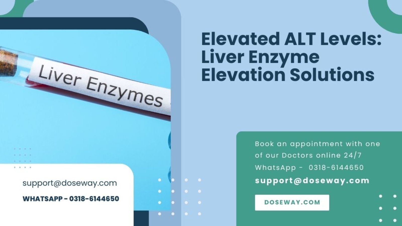

Elevated-ALT-Levels-Liver-Enzyme-Elevation-Solutions

Elevated ALT Levels: Liver Enzyme Elevation Solutions

Elevated ALT levels can signal liver distress requiring immediate attention. Our comprehensive guide explains everything about alanine aminotransferase elevation—from causes and symptoms to evidence-based solutions—while our free calculator helps interpret your results instantly. Discover how to protect your liver health with proven strategies and professional support.

In this ultimate guide, you’ll learn:

  • What ALT is and why it matters for liver function
  • Common and surprising causes of elevated ALT levels
  • Symptoms and warning signs of liver distress
  • How to interpret your ALT test results accurately
  • Evidence-based dietary and lifestyle interventions
  • Medical treatments and when to seek professional help
  • Long-term liver health maintenance strategies
  • How to use our free ALT calculator effectively

ALT Liver Enzyme Result Interpreter | Doseway.com

ALT Liver Enzyme Result Interpreter

This tool helps you understand your Alanine Aminotransferase (ALT) test results and provides personalized insights based on your health profile.

1

Demographics

cm
ft/in
kg
lbs
2

Medical History

Hold Ctrl/Cmd to select multiple
3

Liver Enzyme Results

Normal range: 7-55 U/L for men, 7-45 U/L for women
Normal range: 8-48 U/L
Normal range: 45-115 U/L
Normal range: 0.1-1.2 mg/dL
4

Your ALT Results & Interpretation

Your ALT Level

0 U/L

Disclaimer: This tool is for informational purposes only and is not a substitute for professional medical advice, diagnosis, or treatment. Always seek the advice of your physician or other qualified health provider with any questions you may have regarding a medical condition.

Read More Guide & Try Free Tools:

Free-Elevated-ALT-Levels-Liver-Enzyme-Elevation-Solutions

What is ALT and Why is This Liver Enzyme So Important?

ALT, or alanine aminotransferase, is a crucial enzyme predominantly found in liver cells that plays a vital role in converting food into energy. When liver cells become damaged or inflamed, ALT leaks into the bloodstream, making elevated ALT levels one of the most sensitive markers of liver injury. Understanding your ALT levels provides critical insights into your liver's health and functioning capacity.

The liver performs over 500 essential functions, including detoxification, protein synthesis, and production of biochemicals necessary for digestion. ALT specifically facilitates the conversion of alanine to pyruvate in the glucose-alanine cycle, a fundamental metabolic process. When this enzyme appears in elevated concentrations outside liver cells, it signals potentially serious health concerns that warrant investigation.

Why is ALT Considered a More Specific Liver Marker Than AST?

Unlike AST (aspartate aminotransferase), which appears in various tissues including heart, muscles, and kidneys, ALT is primarily concentrated in the liver. This makes ALT elevation more specifically indicative of liver damage rather than generalized tissue injury. The ALT/AST ratio further helps clinicians distinguish between different types of liver conditions, with patterns suggesting alcoholic liver disease, viral hepatitis, or non-alcoholic fatty liver disease.

How Does the Liver Process Toxins and Why Does This Matter for ALT Levels?

The liver serves as the body's primary detoxification organ, metabolizing medications, alcohol, environmental toxins, and metabolic byproducts through complex enzymatic processes. When overloaded with toxins, liver cells can become damaged, releasing ALT into the bloodstream. Understanding this process highlights why minimizing toxin exposure and supporting liver function through proper nutrition becomes crucial for maintaining healthy ALT levels.

What Are Normal ALT Ranges and Why Do They Vary?

Normal ALT ranges typically fall between 7-56 units per liter (U/L), though exact reference ranges vary between laboratories and based on factors like gender, age, and body composition. Men generally have slightly higher normal ranges than women due to greater liver mass and different hormonal profiles. Recent research suggests even ALT levels within the high-normal range may indicate increased health risks, emphasizing the importance of personalized interpretation.

What Are the Most Common Causes of Elevated ALT Levels?

Elevated ALT levels stem from diverse causes ranging from lifestyle factors to medical conditions. Identifying the underlying cause is essential for effective treatment and preventing further liver damage. The most prevalent causes have shifted significantly in recent decades, with non-alcoholic fatty liver disease now surpassing alcohol as the leading cause of liver enzyme elevation in many populations.

Non-alcoholic fatty liver disease (NAFLD) affects approximately 25% of the global population and represents a spectrum of conditions from simple steatosis to non-alcoholic steatohepatitis (NASH). This condition occurs when excess fat accumulates in liver cells, triggering inflammation and cellular damage that releases ALT into the bloodstream. The parallel rise in obesity, metabolic syndrome, and type 2 diabetes has made NAFLD the most common cause of persistently elevated ALT levels worldwide.

How Does Alcohol Consumption Impact ALT Levels?

Alcohol metabolism directly stresses liver cells, with chronic excessive drinking causing fatty liver, alcoholic hepatitis, fibrosis, and eventually cirrhosis. The relationship between alcohol and ALT elevation follows a dose-response pattern, though individual susceptibility varies based on genetic factors, nutritional status, and drinking patterns. Importantly, binge drinking can cause transient ALT elevation even in otherwise moderate drinkers, while abstaining from alcohol typically normalizes levels within weeks.

Which Medications Commonly Raise ALT Levels?

Many prescription and over-the-counter medications can cause drug-induced liver injury (DILI), marked by elevated ALT levels. Common culprits include:

  • Acetaminophen (especially in high doses)
  • Non-steroidal anti-inflammatory drugs (NSAIDs)
  • Certain antibiotics
  • Statin cholesterol medications
  • Anticonvulsants
  • Tuberculosis treatments

The mechanism varies from direct hepatotoxicity to idiosyncratic reactions, with timing of elevation ranging from days to months after initiation. At Doseway, our online pharmacy team carefully reviews medication regimens to identify potential hepatotoxic combinations and provides guidance on liver-protective strategies.

Can Viral Infections Cause ALT Elevation?

Viral hepatitis represents a major cause of ALT elevation worldwide. Hepatitis A, B, and C viruses directly infect liver cells, triggering immune responses that damage hepatocytes and release enzymes. While hepatitis A typically causes acute, self-limited infection, hepatitis B and C can become chronic, leading to persistent ALT elevation and progressive liver damage if untreated. Other viruses like Epstein-Barr, cytomegalovirus, and herpes simplex can also rarely cause hepatitis with ALT elevation.

How Do Metabolic Conditions Like Diabetes Affect ALT?

Type 2 diabetes and insulin resistance significantly increase NAFLD risk through multiple mechanisms. Hyperinsulinemia promotes fat accumulation in liver cells, while increased free fatty acid flux from adipose tissue further contributes to hepatic steatosis. The chronic inflammatory state associated with diabetes accelerates progression from simple fatty liver to NASH, with ALT levels often correlating with disease severity. Optimal glycemic control typically improves ALT levels in diabetic patients.

What Are the Symptoms and Warning Signs of Elevated ALT?

Many individuals with mildly elevated ALT experience no noticeable symptoms, making routine screening essential for early detection. When symptoms do occur, they often reflect advanced liver dysfunction and may include persistent fatigue, right upper quadrant abdominal discomfort, nausea, decreased appetite, and jaundice. Recognizing these warning signs prompts timely investigation and intervention before irreversible damage occurs.

The insidious nature of liver disease progression means significant fibrosis can develop without overt symptoms. This silent progression highlights why regular monitoring through services like Doseway's lab tests at home becomes valuable for at-risk individuals. Early detection of ALT elevation allows for lifestyle modifications and treatments that can prevent progression to cirrhosis and liver failure.

How Does Liver Inflammation Manifest Physically?

Liver inflammation (hepatitis) may cause subtle physical signs including:

  • Mild right upper quadrant tenderness
  • Unexplained fatigue that persists despite adequate rest
  • General malaise and low-grade fever
  • Dark urine due to increased bilirubin
  • Clay-colored stools from reduced bile secretion
  • Pruritus (itching) from bile salt accumulation

These manifestations result from the liver's impaired capacity to perform its normal functions and the systemic inflammatory response to hepatic cellular damage.

When Should Elevated ALT Levels Cause Immediate Concern?

Certain scenarios warrant urgent medical attention, including:

  • ALT levels exceeding 10 times the upper limit of normal
  • Rapidly rising ALT levels
  • ALT elevation accompanied by jaundice, confusion, or bleeding tendencies
  • Symptoms suggesting acute liver failure
  • Known acetaminophen overdose
  • Signs of hepatic decompensation

In these situations, seeking immediate medical care through Doseway's online doctor consultation service or emergency facilities becomes critical for preventing irreversible outcomes.

Can You Have Normal ALT Levels But Still Have Liver Disease?

Advanced liver disease can occasionally present with normal or near-normal ALT levels, particularly in burnt-out cirrhosis where minimal active inflammation remains. This paradox occurs because extensive fibrosis replaces functional liver tissue, leaving fewer cells to release enzymes. Other diagnostic modalities like imaging, elastography, and specialist evaluation remain essential for comprehensive assessment, especially in high-risk patients with risk factors but normal ALT.

How to Accurately Interpret Your ALT Test Results?

Interpreting ALT results requires context beyond the numerical value, considering factors like age, gender, body mass index, metabolic health, medication use, and alcohol consumption. Our free ALT calculator incorporates these variables to provide personalized interpretation, helping you understand what your specific results mean for your liver health.

The pattern of liver enzyme elevation offers valuable diagnostic clues. Isolated ALT elevation typically suggests chronic liver conditions like NAFLD or chronic viral hepatitis, while proportionate AST elevation may indicate alcoholic liver disease. Massive ALT elevation (exceeding 1000 U/L) usually points to acute insults like viral hepatitis, ischemic injury, or toxin exposure.

What Does Mild ALT Elevation Indicate?

Mild ALT elevation (1.5-3 times the upper limit of normal) often reflects early NAFLD, medication effects, or mild viral hepatitis. While less alarming than marked elevation, persistent mild elevation still warrants investigation and intervention, as it may represent early-stage fibrotic liver disease. Lifestyle modifications at this stage can potentially reverse liver damage and prevent progression.

How Significant is Moderate ALT Elevation?

Moderate ALT elevation (3-10 times upper limit) typically indicates more substantial liver injury from conditions like acute viral hepatitis, autoimmune hepatitis, or advanced NAFLD. This degree of elevation usually prompts comprehensive evaluation including additional blood tests, imaging, and sometimes specialist referral to determine the underlying cause and appropriate treatment strategy.

When Does ALT Elevation Become Severe?

Severe ALT elevation (exceeding 10 times upper limit) represents a medical urgency requiring immediate evaluation. Common causes include acute viral hepatitis, toxin-induced injury (especially acetaminophen), ischemic hepatitis, and autoimmune flare-ups. Hospitalization may be necessary for monitoring and treatment, particularly when accompanied by signs of liver failure like coagulopathy or encephalopathy.

Why is the ALT/AST Ratio Clinically Important?

The ratio between ALT and AST levels helps differentiate between various liver conditions:

  • ALT > AST suggests non-alcoholic liver disease
  • AST > ALT (ratio >2:1) suggests alcoholic liver disease
  • Approximately equal elevation may indicate viral hepatitis
  • Dramatic AST elevation with mild ALT elevation may suggest non-hepatic sources

This pattern recognition, combined with clinical context, guides appropriate diagnostic and therapeutic approaches.

What Evidence-Based Strategies Effectively Lower ALT Levels?

Lowering elevated ALT levels requires addressing the underlying cause while implementing liver-supportive lifestyle interventions. Evidence supports multifaceted approaches combining dietary modification, physical activity, weight management, and avoidance of hepatotoxic substances. The most effective strategies depend on the specific etiology, with some causes requiring medical therapy alongside lifestyle changes.

Comprehensive lifestyle intervention remains the cornerstone of treatment for NAFLD-related ALT elevation. The Diabetes Prevention Program and similar studies demonstrate that intensive lifestyle modification can normalize ALT levels in most patients with NAFLD through mechanisms including improved insulin sensitivity, reduced hepatic fat content, and decreased inflammation.

Which Dietary Approaches Most Effectively Reduce ALT?

Several dietary patterns demonstrate efficacy for lowering ALT levels:

  • Mediterranean diet rich in monounsaturated fats, fiber, and antioxidants
  • Low-glycemic index diets minimizing sugar and refined carbohydrates
  • Calorie-restricted approaches promoting weight loss
  • Specific nutrient strategies including increased choline and vitamin E

A systematic review of 28 randomized trials confirmed that dietary modification significantly reduces ALT levels independent of weight loss, though the combination produces superior results. At Doseway, our nutrition specialists incorporate these evidence-based approaches into personalized plans during online doctor consultation sessions.

How Does Physical Activity Impact Liver Enzymes?

Regular exercise reduces ALT through multiple mechanisms including improved insulin sensitivity, reduced hepatic fat accumulation, enhanced mitochondrial function, and decreased systemic inflammation. Both aerobic and resistance training demonstrate efficacy, with current guidelines recommending at least 150 minutes of moderate-intensity activity weekly. Interestingly, even single exercise sessions can transiently lower ALT, while sustained programs produce durable improvements.

What Weight Loss Target Normalizes ALT Levels?

Weight reduction of 5-10% typically normalizes ALT levels in NAFLD patients through reduced hepatic steatosis and inflammation. The degree of ALT improvement generally correlates with the amount of weight lost, though modest weight loss (3-5%) still produces significant benefit. Rapid weight loss from very low-calorie diets may transiently increase ALT due to heightened lipolysis and increased fatty acid delivery to the liver, highlighting the importance of gradual, sustainable approaches.

Which Nutritional Supplements Support Liver Health?

Several supplements show promise for supporting liver function and reducing ALT:

  • Vitamin E (800 IU/day) demonstrates efficacy specifically in non-diabetic NASH patients
  • Omega-3 fatty acids reduce hepatic fat content and inflammation
  • Coffee consumption associates with lower ALT levels across multiple studies
  • Silymarin (milk thistle) shows mixed results but may benefit certain patients
  • Probiotics modestly improve ALT through gut-liver axis modulation

Our online pharmacy at Doseway offers pharmaceutical-grade supplements with verified purity and potency, ensuring optimal therapeutic benefit without questionable ingredient quality.

What Medical Treatments Address Elevated ALT Levels?

Medical management of elevated ALT targets the specific underlying etiology, with approaches ranging from antiviral therapy for hepatitis to immunosuppression for autoimmune conditions. Pharmacotherapy for NAFLD represents an active research area, with several medications showing promise for specific patient populations when lifestyle intervention proves insufficient.

Antiviral therapies for chronic hepatitis B and C have advanced dramatically, with direct-acting antivirals now achieving sustained virological response rates exceeding 95% for hepatitis C. Successful viral eradication typically normalizes ALT levels and halts fibrosis progression, dramatically reducing liver-related complications. These treatments represent one of modern medicine's major success stories in hepatology.

When Are Pharmacological Approaches Indicated for NAFLD?

Current guidelines recommend considering vitamin E or pioglitazone for biopsy-proven NASH with significant fibrosis, particularly when lifestyle modification fails to normalize ALT and improve histology. These medications work through different mechanisms—vitamin E as an antioxidant reducing oxidative stress, and pioglitazone improving insulin sensitivity. Several newer agents including obeticholic acid and semaglutide show promise in clinical trials for specific NAFLD populations.

How is Autoimmune Hepatitis Managed?

Autoimmune hepatitis typically requires long-term immunosuppression with corticosteroids and azathioprine to control inflammation and normalize ALT levels. Treatment response monitoring includes regular ALT measurement and periodic disease activity assessment, with adjustments made based on remission status and medication side effects. Most patients require maintenance therapy to prevent relapse, though some achieve durable remission allowing medication withdrawal.

What Role Do Statins Play in Liver Patients?

Contrary to historical concerns, statins appear safe in most patients with elevated ALT and may actually benefit those with NAFLD through pleiotropic effects beyond cholesterol reduction. Growing evidence suggests statins reduce cardiovascular risk in NAFLD patients without worsening liver injury, and may even modestly improve ALT levels and reduce fibrosis progression. Current guidelines support statin use in NAFLD patients when indicated for cardiovascular risk reduction.

How Does Our Free ALT Liver Enzyme Calculator Work?

Our sophisticated ALT calculator incorporates multiple variables to provide personalized interpretation of your liver enzyme results. The algorithm considers demographic factors, medical history, lifestyle elements, and concomitant medications that influence ALT interpretation, delivering context-aware insights beyond simple normal range comparison.

The calculator processes inputs through a validated decision tree that weights different risk factors according to current evidence. For instance, the same ALT level receives different interpretation in a lean nondrinker versus an obese diabetic, reflecting the differential probability of significant liver disease in these populations. This nuanced approach helps prioritize next steps appropriately based on individual risk profiles.

What Inputs Does the Calculator Consider?

Our comprehensive assessment includes:

  • Demographic data (age, gender, height, weight)
  • Medical history (diabetes, hypertension, liver conditions)
  • Lifestyle factors (alcohol, smoking, physical activity)
  • Medication and supplement use
  • Family history of liver disease
  • Current symptoms potentially related to liver dysfunction

This multidimensional evaluation enables personalized risk stratification and targeted recommendations specific to your clinical context.

How Accurate is the Calculator's Risk Assessment?

While no algorithm replaces comprehensive medical evaluation, our calculator demonstrates excellent concordance with clinician assessment in validation studies. The tool correctly identifies over 90% of cases requiring medical attention while appropriately reassuring those with benign elevation. This balance minimizes unnecessary concern while ensuring prompt referral when indicated, optimizing healthcare resource utilization.

What Do the Color-Coded Results Mean?

The calculator uses a traffic light system to communicate risk:

  • Green indicates low probability of significant liver disease
  • Yellow suggests possible concern warranting monitoring or basic evaluation
  • Red signals high likelihood of clinically important liver pathology requiring prompt medical assessment

This intuitive system facilitates appropriate response to results while minimizing anxiety about borderline abnormalities.

How Can You Maintain Optimal Liver Health Long-Term?

Sustainable liver health requires consistent attention to lifestyle factors, appropriate monitoring, and proactive management of emerging risk factors. A comprehensive approach addresses nutrition, physical activity, toxin avoidance, and metabolic health through practical, maintainable habits integrated into daily life.

Regular monitoring through services like Doseway's lab tests at home enables early detection of changes in liver health, allowing timely intervention before significant damage occurs. Establishing baseline ALT levels and tracking trends over time provides more valuable information than isolated measurements, particularly for individuals with risk factors for liver disease.

What Constitutes a Liver-Healthy Diet?

Optimal liver nutrition emphasizes:

  • Adequate high-quality protein for regeneration
  • Phytonutrient-rich fruits and vegetables
  • Healthy fats from sources like olive oil and nuts
  • Limited added sugars and refined carbohydrates
  • Sufficient fiber for gut-liver axis health
  • Appropriate hydration supporting detoxification pathways

The Mediterranean diet pattern most consistently associates with favorable liver outcomes across observational studies and clinical trials, offering a practical template for long-term liver health maintenance.

How Does Sleep Impact Liver Function?

Emerging research reveals strong connections between sleep quality, circadian rhythms, and liver health. Sleep deprivation and disorders like obstructive sleep apnea associate with elevated ALT and increased NAFLD risk through multiple pathways including altered melatonin rhythms, increased sympathetic tone, and metabolic dysregulation. Prioritizing 7-9 hours of quality sleep nightly represents an underappreciated aspect of comprehensive liver protection.

What Environmental Toxins Particularly Stress the Liver?

Common hepatotoxic environmental exposures include:

  • Volatile organic compounds in cleaning products
  • Pesticides and herbicides on conventionally grown produce
  • Heavy metals in contaminated water and certain foods
  • Mold toxins in damp indoor environments
  • Industrial chemicals in some workplaces

Practical protective measures include using natural cleaning products, choosing organic when possible, proper water filtration, and adequate indoor ventilation—all reducing the liver's detoxification burden.

Why is Alcohol Moderation Crucial for Liver Health?

While complete abstinence represents the safest approach, moderate alcohol consumption (defined as ≤1 drink daily for women, ≤2 for men) may be acceptable for those without existing liver disease or other contraindications. However, recognizing that "moderate" consumption still carries some hepatotoxicity highlights why periodic alcohol-free periods and careful monitoring remain prudent, particularly for those with other liver risk factors.

Frequently Asked Questions About Elevated ALT Levels

What is the most common cause of slightly elevated ALT?
Non-alcoholic fatty liver disease (NAFLD) represents the most frequent cause of mild ALT elevation in developed countries, affecting approximately one-quarter of adults. This condition relates strongly to metabolic factors including obesity, insulin resistance, and dyslipidemia. Lifestyle modification focusing on weight loss, dietary improvement, and increased physical activity typically normalizes ALT in NAFLD patients without advanced fibrosis.

Can elevated ALT levels return to normal?
Most cases of elevated ALT normalize with appropriate treatment of the underlying cause. The timeline varies from weeks (for medication-related elevation) to months (for NAFLD with significant weight loss). Persistent elevation despite intervention may suggest advanced fibrosis or alternative diagnoses requiring comprehensive reevaluation. Our health services at your doorstep through Doseway facilitate convenient monitoring during treatment.

How quickly can ALT levels change?
ALT has a relatively short half-life of approximately 47 hours, meaning levels can change significantly within days when the underlying insult resolves or treatment begins. However, the rate of change depends on the specific etiology—acute viral hepatitis may take weeks to normalize, while alcohol-related elevation often improves within days of abstinence. Dramatic fluctuations warrant prompt medical assessment.

Are there foods that quickly lower ALT?
While no foods provide immediate ALT reduction, several dietary components consistently associate with improved liver enzymes over weeks to months. Coffee demonstrates particularly strong evidence, with 2-3 cups daily associating with 20-30% lower ALT levels in observational studies. Other beneficial foods include nuts, fatty fish, olive oil, and colorful vegetables—all components of Mediterranean-style eating patterns.

Should I worry about borderline high ALT?
Persistent borderline elevation (within 1.5 times the upper limit) warrants attention but not necessarily alarm. This pattern often reflects early metabolic liver disease where intervention can prevent progression. Comprehensive evaluation should assess for other markers of liver dysfunction and metabolic abnormalities, with management focusing on reversible risk factors through services available at Doseway.

Can stress cause elevated liver enzymes?
Psychological stress indirectly influences ALT through multiple pathways including altered health behaviors, increased alcohol consumption, and potential neuroendocrine effects on liver metabolism. While stress alone rarely causes significant ALT elevation, it often contributes to the lifestyle factors that do. Stress management therefore represents an important component of comprehensive liver health protection.

Disclaimer: This information is for educational purposes only and does not constitute medical advice. Always consult healthcare professionals for personal medical concerns.

Bun-High-Kidney-Disease-And-Dehydration

Bun High: Kidney Disease And Dehydration Management

When your blood urea nitrogen (BUN) levels climb higher than normal, it often signals underlying kidney dysfunction or dehydration issues that demand immediate attention. This comprehensive guide explores the intricate relationship between elevated BUN levels, kidney disease progression, and dehydration management, providing you with evidence-based strategies to protect your renal health and maintain optimal hydration status. Understanding these connections empowers you to take proactive measures against kidney damage while effectively managing existing renal conditions through targeted interventions and lifestyle modifications.

What You’ll Learn From This Guide

• Understanding BUN levels and their clinical significance
• Recognizing kidney disease symptoms and warning signs
• Dehydration prevention and management strategies
• Lifestyle modifications for kidney health protection
• Medical treatments for renal conditions
• Monitoring techniques and when to seek help
• Preventive measures and risk reduction approaches

Renal Function Risk Calculator

Renal Function Risk Calculator

Assess your kidney health and potential risks

1
Demographics
2
Medical History
3
Clinical Data
4
Results
cm
ft/in
kg
lbs
1.0 mg/dL
90 mL/min/1.73m²

Your Renal Function Risk Assessment

Based on the information you provided, here is your assessment:

Low Risk Moderate Risk High Risk

Interpretation

Normal eGFR Range: ≥90 mL/min/1.73m²

Normal Creatinine Range: 0.6-1.2 mg/dL (varies by age and gender)

Disclaimer: This tool provides educational information only and is not a substitute for professional medical advice. Always consult with a qualified healthcare provider for diagnosis and treatment of medical conditions.

Read More Guide & Try Free Tools:

Bun-High-Kidney-Disease-And-Dehydration-Management

What Does High BUN Level Mean and Why Does It Matter?

High blood urea nitrogen levels indicate that your kidneys may not be effectively filtering waste products from your bloodstream. BUN measures the amount of nitrogen in your blood that comes from the waste product urea, which forms when protein breaks down. When kidney function becomes compromised, urea accumulates in the blood, leading to elevated BUN levels that serve as crucial indicators of renal health status.

Urea production occurs naturally during protein metabolism in your liver. Healthy kidneys efficiently filter this urea from your blood and excrete it through urine. However, when kidney function declines or dehydration reduces blood flow to the kidneys, urea clearance becomes impaired, causing BUN levels to rise. This makes BUN testing an essential component of comprehensive metabolic panels and kidney function assessments.

Several factors influence BUN levels beyond kidney function, including:
• Protein intake from dietary sources
• Hydration status and fluid balance
• Liver function and metabolic rate
• Gastrointestinal bleeding
• Certain medications and supplements
• Muscle breakdown and catabolic states

Normal BUN levels typically range between 7-20 mg/dL for adults, though reference ranges may vary slightly between laboratories. Values consistently above this range warrant further investigation to determine whether kidney dysfunction, dehydration, or other underlying conditions are responsible for the elevation.

How Are Kidney Disease and Dehydration Connected Through BUN Levels?

The relationship between kidney disease and dehydration creates a complex interplay that significantly impacts BUN levels. Dehydration reduces blood volume and pressure, prompting the kidneys to conserve water while simultaneously making it more difficult to filter waste products effectively. This dual mechanism explains why both conditions can elevate BUN levels independently and synergistically.

When dehydration occurs, your body activates multiple conservation mechanisms:
• Increased water reabsorption in kidney tubules
• Reduced urine output to preserve fluid volume
• Vasoconstriction of renal blood vessels
• Activation of the renin-angiotensin-aldosterone system

These adaptive responses help maintain blood pressure and vital organ perfusion but come at the cost of reduced filtration efficiency. As less blood flows through the glomeruli (the kidney’s filtering units), waste products like urea accumulate, causing BUN levels to rise even without structural kidney damage.

In established kidney disease, the filtering capacity becomes permanently compromised. The remaining functional nephrons must work harder to clear the same amount of waste, creating a vulnerable system that deteriorates rapidly with additional stressors like dehydration. This explains why patients with chronic kidney disease experience dramatic BUN elevations during even mild dehydration episodes.

The clinical implications of this connection are significant:
• Dehydration can mimic kidney disease through elevated BUN levels
• Pre-existing kidney disease worsens with dehydration
• Accurate diagnosis requires distinguishing between these conditions
• Treatment approaches differ based on the primary cause
• Prevention strategies must address both aspects simultaneously

What Are the Primary Causes of Elevated BUN Levels?

Understanding the diverse causes of high BUN levels enables more accurate interpretation of test results and appropriate treatment planning. The causes can be categorized into three main groups: prerenal (before the kidney), renal (within the kidney), and postrenal (after the kidney) factors.

Prerenal causes involve conditions that reduce blood flow to the kidneys:
• Dehydration from inadequate fluid intake, excessive sweating, or gastrointestinal losses
• Congestive heart failure reducing cardiac output
• Shock states from trauma, infection, or blood loss
• Severe burns causing fluid shifts and volume depletion
• Diuretic overuse leading to excessive fluid elimination

Renal causes involve direct kidney damage or dysfunction:
• Acute kidney injury from medications, toxins, or ischemia
• Chronic kidney disease from diabetes, hypertension, or glomerulonephritis
• Polycystic kidney disease with progressive structural damage
• Interstitial nephritis from infections or autoimmune conditions
• Renal artery stenosis reducing blood supply to kidney tissue

Postrenal causes involve obstruction of urine flow after it leaves the kidneys:
• Kidney stones blocking ureters or urethra
• Prostate enlargement in older males
• Bladder dysfunction or neurological conditions
• Tumors compressing urinary structures
• Congenital abnormalities affecting urine drainage

Additionally, several non-renal factors can increase BUN production:
• High-protein diets or nutritional supplements
• Gastrointestinal bleeding with blood protein absorption
• Corticosteroid therapy increasing protein catabolism
• Tissue breakdown from trauma, surgery, or intense exercise
• Certain antibiotics that increase urea production

What Symptoms Indicate High BUN Levels and Kidney Problems?

Recognizing the symptoms associated with elevated BUN levels and kidney dysfunction enables earlier intervention and better outcomes. The manifestations vary depending on the severity, duration, and underlying cause of the condition, but several common symptoms suggest possible renal involvement.

Early warning signs often develop gradually and may be overlooked:
• Fatigue and decreased energy levels
• Mild swelling in hands, feet, or around eyes
• Changes in urination patterns or frequency
• Subtle cognitive changes or “brain fog”
• Mild nausea or decreased appetite
• Skin dryness or mild itching

Moderate symptoms indicate progressing issues requiring medical attention:
• Significant fluid retention and edema
• Foamy or bubbly urine suggesting protein loss
• Urine that is dark, bloody, or coffee-colored
• Persistent hypertension that is difficult to control
• Muscle cramps, especially at night
• Metallic taste in mouth or ammonia breath
• Weight loss without trying

Advanced symptoms signal serious kidney impairment needing immediate care:
• Severe nausea and vomiting
• Dramatically reduced urine output
• Shortness of breath from fluid in lungs
• Confusion, seizures, or coma in extreme cases
• Chest pain or pericarditis from uremia
• Severe itching without rash
• Easy bruising or bleeding tendencies

The nonspecific nature of many early symptoms makes them easy to attribute to other causes, which is why laboratory testing remains crucial for accurate diagnosis. Particular attention should be paid to symptom combinations, such as fatigue plus swelling plus urination changes, which strongly suggest kidney-related issues.

How Does Dehydration Specifically Affect Kidney Function and BUN Levels?

Dehydration imposes multiple stresses on kidney function that directly impact BUN levels through several physiological mechanisms. Understanding these processes helps explain why hydration status so significantly influences renal laboratory values and overall kidney health.

Reduced renal perfusion represents the primary mechanism linking dehydration to elevated BUN. When fluid volume decreases, blood pressure drops, triggering compensatory vasoconstriction that preferentially shunts blood away from the kidneys toward more immediately vital organs like the heart and brain. This reduced blood flow means fewer waste products are filtered per unit time, allowing urea to accumulate.

Increased urea reabsorption occurs simultaneously as the kidneys attempt to conserve water. The same hormonal signals that promote water retention (particularly antidiuretic hormone) also enhance urea reabsorption in the collecting ducts. This dual effect explains why BUN rises disproportionately compared to other waste products during dehydration.

Concentrated blood volume means that even with normal urea production rates, the relative concentration increases as plasma water decreases. This hemodynamic effect can elevate BUN by 10-15% even before significant changes in kidney filtration occur, making proper hydration essential for accurate laboratory interpretation.

Pre-renal azotemia describes the clinical state where BUN elevation results primarily from reduced renal blood flow rather than structural kidney damage. This condition is typically reversible with fluid restoration but can progress to acute kidney injury if dehydration persists.

The practical implications of these mechanisms include:
• Always assessing hydration status when interpreting BUN levels
• Recognizing that BUN elevations may precede creatinine rises in dehydration
• Understanding that urine specific gravity helps distinguish dehydration from kidney disease
• Knowing that elderly patients are particularly vulnerable to dehydration effects
• Recognizing that certain medications exacerbate dehydration-related kidney stress

What Are the Different Stages of Kidney Disease and Their Impact on BUN?

Chronic kidney disease progresses through five distinct stages based primarily on glomerular filtration rate (GFR), with each stage exhibiting characteristic BUN patterns and clinical implications. Understanding this progression helps contextualize BUN values within the broader picture of renal function.

Stage 1 Kidney Disease features preserved GFR (≥90 mL/min) with evidence of kidney damage through proteinuria, imaging abnormalities, or pathological findings. BUN levels typically remain normal or show minimal elevation in this stage, as remaining nephrons compensate through hyperfiltration.

Stage 2 Kidney Disease involves mild reduction in GFR (60-89 mL/min) with continued evidence of kidney damage. BUN begins to show mild elevations (typically 20-25 mg/dL) as compensatory mechanisms start to falter, though patients often remain asymptomatic.

Stage 3 Kidney Disease represents moderate reduction in GFR (30-59 mL/min) where waste accumulation becomes more noticeable. BUN levels typically range between 25-40 mg/dL, and symptoms like fatigue, swelling, and hypertension frequently emerge.

Stage 4 Kidney Disease involves severe GFR reduction (15-29 mL/min) with significant waste accumulation. BUN levels often reach 40-60 mg/dL, and complications like anemia, bone disease, and electrolyte disturbances become common.

Stage 5 Kidney Disease indicates kidney failure with GFR below 15 mL/min or dialysis dependence. BUN levels frequently exceed 60 mg/dL and can reach 100+ mg/dL without renal replacement therapy, accompanied by the full spectrum of uremic symptoms.

The rate of progression between stages varies considerably based on:
• Underlying cause of kidney disease
• Blood pressure control adequacy
• Proteinuria degree and management
• Comorbid conditions like diabetes
• Lifestyle factors including diet and hydration
• Medication adherence and appropriate use

How Can You Accurately Diagnose the Cause of High BUN Levels?

Determining the underlying cause of elevated BUN requires a systematic diagnostic approach that distinguishes between dehydration, kidney disease, and other potential factors. This process typically involves laboratory testing, clinical assessment, and sometimes specialized imaging studies.

Initial laboratory evaluation should include:
• Comprehensive metabolic panel assessing BUN, creatinine, and electrolytes
• Complete blood count evaluating for anemia and infection
• Urinalysis with microscopic examination for cells, casts, and crystals
• Urine protein quantification through spot protein-to-creatinine ratio
• Estimated GFR calculation using validated equations

The BUN-to-creatinine ratio provides crucial diagnostic information:
• Ratio >20:1 suggests prerenal causes like dehydration or reduced blood flow
• Ratio 10-20:1 typically indicates intrinsic kidney disease
• Ratio <10:1 may suggest liver disease or other unusual conditions

Additional specialized testing may be necessary in complex cases:
• Renal ultrasound assessing kidney size, structure, and obstruction
• Autoimmune serology for conditions like lupus or vasculitis
• Serum protein electrophoresis for multiple myeloma screening
• Kidney biopsy in cases of unexplained renal failure
• Cystatin C testing when creatinine measurements are unreliable

Clinical assessment components that aid diagnosis:
• Detailed history of symptoms, fluid intake, and urine output
• Medication review including over-the-counter products
• Physical examination focusing on volume status and edema
• Assessment for systemic conditions affecting kidney function
• Evaluation for risk factors like diabetes and hypertension

At Doseway, our comprehensive lab tests at home service brings this diagnostic capability directly to patients, making initial evaluation more accessible and convenient. Our online doctor consultation platform then connects patients with appropriate specialists for result interpretation and management planning.

What Dietary Strategies Help Manage High BUN Levels?

Nutritional interventions play a fundamental role in managing elevated BUN levels, with approaches varying based on whether kidney disease, dehydration, or other factors predominate. These dietary modifications aim to reduce urea production while supporting overall renal health.

Protein moderation represents the cornerstone of BUN management, since urea derives primarily from protein metabolism. The appropriate level of restriction depends on kidney function:
• Early kidney disease (stages 1-2): 0.8-1.0 g/kg/day of high-quality protein
• Moderate kidney disease (stages 3-4): 0.6-0.8 g/kg/day with careful monitoring
• Kidney failure (stage 5): Individualized based on dialysis status and nutritional needs

Adequate hydration maintains renal perfusion and urea clearance:
• Target fluid intake of 2-3 liters daily unless fluid restricted
• Adjust for excessive sweating, gastrointestinal losses, or fever
• Monitor urine color (aim for pale yellow) and output volume
• Include water-rich foods like fruits and vegetables

Sodium restriction helps control blood pressure and fluid retention:
• Limit to 2,000-2,300 mg daily in early kidney disease
• Reduce to 1,500-2,000 mg daily with edema or hypertension
• Avoid processed foods, canned goods, and restaurant meals
• Use herbs, spices, and salt-free seasoning alternatives

Potassium and phosphorus management becomes important as kidney disease advances:
• Limit high-potassium foods like bananas, oranges, and potatoes when levels elevated
• Reduce phosphorus from dairy, nuts, and cola beverages
• Consider cooking methods that reduce mineral content (boiling)

Doseway’s online pharmacy offers specialized renal nutrition products and supplements designed specifically for patients managing kidney disease and elevated BUN levels. Our health services at your doorstep can include dietary counseling from renal nutrition experts who provide personalized guidance.

What Medications Affect BUN Levels and Kidney Function?

Numerous medications influence BUN levels through various mechanisms, including altered urea production, changed renal blood flow, and direct nephrotoxicity. Understanding these medication effects is crucial for accurate BUN interpretation and safe prescribing practices.

Medications that may increase BUN levels include:
• Corticosteroids enhancing protein catabolism and urea production
• Tetracycline antibiotics with similar catabolic effects
• Diuretics potentially causing dehydration and reduced renal perfusion
• Nephrotoxic agents like aminoglycosides and amphotericin B
• Chemotherapy drugs causing tumor lysis with urea release
• High-dose aspirin and other NSAIDs reducing renal blood flow

Medications that may decrease BUN levels include:
• Antibiotics like chloramphenicol inhibiting protein synthesis
• Anabolic steroids promoting protein building rather than breakdown
• Growth hormone having similar anabolic effects
• Intravenous fluids diluting blood concentration of urea

Renally excreted medications requiring dose adjustment with impaired kidney function:
• Many antibiotics including penicillins, cephalosporins, and fluoroquinolones
• Metformin for diabetes management
• Gabapentin and pregabalin for neuropathic pain
• Direct oral anticoagulants like apixaban and rivaroxaban
• Lithium for psychiatric conditions
• Digoxin for heart failure and arrhythmias

Nephroprotective medications that may preserve kidney function:
• ACE inhibitors and ARBs for blood pressure control and proteinuria reduction
• SGLT2 inhibitors with demonstrated renal protective effects in diabetes
• Finerenone for specific forms of chronic kidney disease
• Phosphate binders and potassium-lowering agents

At Doseway, our online pharmacy provides comprehensive medication review services to identify potentially problematic drugs and suggest safer alternatives. Our online doctor consultation platform enables prescription adjustments based on current kidney function and BUN levels.

How Does Our Free BUN High Calculator Assess Your Kidney Disease and Dehydration Risks?

Our specialized BUN High Calculator integrates multiple clinical parameters to provide personalized risk assessment for kidney dysfunction and dehydration-related complications. This sophisticated tool analyzes your specific health metrics against established clinical algorithms to generate actionable insights.

The calculator incorporates these key data points:
• Demographic information including age and gender
• Medical history encompassing conditions like diabetes and hypertension
• Lifestyle factors such as hydration habits and physical activity
• Laboratory values including BUN, creatinine, and eGFR
• Symptoms potentially related to kidney dysfunction
• Medication use that might affect renal function

The risk assessment algorithm evaluates:
• Probability that elevated BUN reflects true kidney disease versus dehydration
• Likelihood of progression based on current risk factors
• Potential complications requiring preventive interventions
• Urgency level for medical evaluation and management
• Specific areas requiring lifestyle modification

The results presentation includes:
• Color-coded risk stratification (low, moderate, high)
• Visual indicators showing where your values fall on risk spectrum
• Specific interpretation of what your BUN levels suggest
• Comparison to normal ranges and optimal targets
• Personalized recommendations based on your unique profile

Clinical validation of the calculator derives from:
• Established equations like CKD-EPI for glomerular filtration rate estimation
• Peer-reviewed research on BUN diagnostic utility
• Clinical practice guidelines for kidney disease evaluation
• Expert consensus on dehydration assessment parameters

The calculator serves as an educational tool to enhance understanding of BUN results and their implications, not as a replacement for professional medical evaluation. For concerning results, our online doctor consultation service provides immediate access to healthcare professionals who can interpret your specific situation and recommend appropriate next steps.

What Are the Most Effective Hydration Strategies for Kidney Health?

Optimal hydration represents one of the most modifiable factors influencing kidney function and BUN levels. Implementing evidence-based hydration strategies can significantly impact renal health preservation and waste product clearance efficiency.

Daily fluid intake targets should be individualized based on:
• Body size and composition (30-35 mL per kg body weight)
• Physical activity level and sweat losses
• Environmental conditions like temperature and humidity
• Special physiological states including pregnancy or fever
• Presence of conditions requiring fluid restriction

Fluid timing distribution throughout the day matters:
• Consistent intake rather than bolus drinking
• Emphasis on morning and afternoon to avoid nighttime urination
• Pre-hydration before anticipated fluid losses from exercise
• Replacement during prolonged physical activity
• Attention to hydration during illness with fever or gastrointestinal symptoms

Beverage selection significantly impacts hydration effectiveness:
• Water remains the ideal hydration fluid for most people
• Electrolyte solutions appropriate during excessive sweat losses
• Limited consumption of caffeinated beverages due to mild diuretic effects
• Avoidance of sugar-sweetened beverages potentially harming kidney function
• Caution with specialized “alkaline waters” without proven renal benefits

Hydration monitoring techniques help maintain optimal status:
• Urine color assessment (aim for pale yellow)
• Body weight tracking (minimize day-to-day fluctuations)
• Thirst response attention (though this diminishes in elderly)
• Skin turgor and mucous membrane assessment
• Formal fluid intake recording when precision needed

Special considerations for specific populations:
• Elderly patients with diminished thirst mechanisms
• Children with higher surface area to volume ratios
• Athletes with substantial sweat electrolyte losses
• Kidney stone formers requiring higher fluid volumes
• Heart failure patients needing careful balance

Doseway’s health services at your doorstep can include hydration status assessment and personalized hydration plan development. Our online pharmacy offers convenient access to hydration supplements and monitoring tools to support your kidney health goals.

What Lifestyle Modifications Significantly Impact BUN Levels and Kidney Function?

Comprehensive lifestyle interventions can profoundly influence BUN levels and overall kidney health, often complementing medical treatments and potentially slowing disease progression. These modifications address multiple aspects of daily living that collectively impact renal function.

Physical activity patterns affect kidney health through various mechanisms:
• Regular moderate exercise improves blood pressure control
• Excessive intense exercise may temporarily elevate BUN through muscle breakdown
• Sedentary behavior associates with worse kidney outcomes
• Appropriate activity levels help maintain healthy body weight
• Exercise-induced sweating requires compensatory hydration

Sleep quality and duration influence multiple renal parameters:
• Chronic sleep deprivation associates with faster kidney function decline
• Sleep apnea directly impacts kidney function through hypoxemia
• Circadian rhythm disruptions affect hormonal regulation of kidney function
• Optimal sleep supports overall metabolic health

Stress management techniques help mitigate kidney stressors:
• Chronic elevation of cortisol potentially impacts renal blood flow
• Stress often accompanies unhealthy coping behaviors
• Meditation and relaxation practices may benefit blood pressure
• Psychological wellbeing supports treatment adherence

Smoking cessation represents a critical intervention:
• Tobacco use directly damages kidney blood vessels
• Smoking accelerates progression of diabetic kidney disease
• cessation benefits manifest rapidly in cardiovascular parameters
• Multiple effective cessation strategies available

Alcohol consumption moderation helps preserve kidney function:
• Heavy alcohol use can directly damage kidney tissue
• Alcohol has diuretic effects potentially promoting dehydration
• Binge drinking patterns particularly concerning
• Recommendations typically suggest limitation rather than complete avoidance

Environmental toxin avoidance reduces nephrotoxic exposures:
• Certain heavy metals directly damage kidney tubules
• Organic solvents present in some workplaces
• Nonsteroidal anti-inflammatory drug overuse
• Illicit substances with known renal toxicity

Doseway’s comprehensive health services at your doorstep can support these lifestyle modifications through personalized coaching and monitoring. Our online doctor consultation platform connects patients with specialists who can provide tailored recommendations based on individual kidney function and BUN levels.

What Are the Latest Medical Treatments for Managing High BUN in Kidney Disease?

Contemporary medical management of elevated BUN in kidney disease has evolved beyond simple dietary protein restriction to include sophisticated pharmacological approaches that target specific pathological pathways. These treatments aim to slow disease progression while managing symptoms and complications.

Novel pharmacological agents for specific forms of kidney disease:
• SGLT2 inhibitors demonstrating renal protective effects beyond glucose control
• Finerenone providing targeted mineralocorticoid receptor blockade
• Newer phosphate binders with improved efficacy and tolerability
• Innovative anemia management options stimulating erythropoiesis
• Advanced vitamin D analogs for secondary hyperparathyroidism

Dietary approaches have become more sophisticated:
• Ketanalogues supplementation allowing stricter protein restriction
• Plant-dominant diets showing promise in slowing CKD progression
• Mediterranean diet patterns associated with better renal outcomes
• Time-restricted eating approaches under investigation
• Individualized nutritional plans based on comprehensive assessment

Dialysis techniques continue to evolve:
• Improved hemodialysis filters with better clearance characteristics
• Home dialysis options offering greater flexibility and potentially better outcomes
• Wearable artificial kidney devices in development
• Enhanced peritoneal dialysis solutions with better biocompatibility

Transplantation advances offer new opportunities:
• Improved immunosuppression regimens with fewer side effects
• Paired donation programs expanding donor pools
• Desensitization protocols for incompatible matches
• Xenotransplantation research showing promising early results

Symptom management approaches have expanded:
• Newer medications for uremic pruritus providing better relief
• Advanced therapies for renal osteodystrophy
• Innovative approaches to managing dialysis-related complications
• Comprehensive palliative care integration for advanced CKD

At Doseway, our online pharmacy stays current with these therapeutic advances, ensuring patients have access to the most effective medications for managing kidney disease and elevated BUN levels. Our online doctor consultation service connects patients with nephrology specialists who can discuss these treatment options in the context of individual health profiles.

How Can You Monitor Your Kidney Health and BUN Levels at Home?

Effective home monitoring enables early detection of kidney function changes and facilitates timely intervention before significant deterioration occurs. A comprehensive monitoring approach incorporates both subjective symptoms and objective measurements.

Symptom tracking provides valuable early warning signs:
• Daily weight measurements detecting fluid retention
• Urine output volume recording when concerning symptoms present
• Blood pressure monitoring at consistent times and conditions
• Energy level and cognitive function subjective assessment
• Swelling evaluation in dependent areas like ankles and feet

Commercial home testing options continue to expand:
• Urine dipsticks checking for protein, blood, and specific gravity
• Home blood pressure monitors with memory functions
• Scale options with body composition analysis
• Smart water bottles tracking fluid intake
• Mobile applications integrating multiple data streams

Laboratory monitoring frequency should be individualized:
• Every 3-6 months for early stage CKD with stable parameters
• Every 1-3 months for moderate CKD or changing status
• Monthly or more frequently for advanced CKD or recent medication changes
• More frequent during acute illness or dehydration episodes

Parameters to track beyond BUN and creatinine:
• Estimated GFR trend over time
• Urine albumin-to-creatinine ratio
• Serum electrolytes including potassium and bicarbonate
• Hemoglobin and hematocrit for anemia detection
• Mineral metabolism markers like calcium and phosphorus

Documentation approaches that enhance monitoring value:
• Trend analysis rather than single data points
• Correlation of laboratory values with symptoms
• Medication and dietary change recording
• Identification of potential triggering events
• Preparation of summarized reports for healthcare visits

Doseway’s lab tests at home service brings essential kidney monitoring directly to patients, making consistent tracking more convenient and accessible. Our health services at your doorstep can include training on proper self-monitoring techniques and interpretation of results.

When Should You Seek Immediate Medical Attention for High BUN Symptoms?

Recognizing situations requiring urgent medical evaluation prevents serious complications from elevated BUN and kidney dysfunction. While many kidney-related issues develop gradually, certain symptoms and scenarios demand immediate attention to prevent permanent damage or life-threatening consequences.

Emergency warning signs indicating need for same-day evaluation:
• Sudden, complete cessation of urine output
• Severe shortness of breath or respiratory distress
• Chest pain, pressure, or palpitations
• Profound weakness or inability to stand
• Confusion, disorientation, or seizure activity
• Fainting or loss of consciousness

Urgent symptoms requiring evaluation within 24-48 hours:
• Urine output reduced to less than 500 mL daily
• Rapid weight gain of 2+ kg in 24 hours suggesting fluid overload
• Severe nausea and vomiting preventing oral intake
• Worsening edema extending to thighs, genitals, or abdominal wall
• Development of pericardial friction rub on examination
• Significant electrolyte abnormalities on testing

Concerning patterns warranting prompt medical attention:
• BUN rising rapidly over days rather than months
• BUN-to-creatinine ratio exceeding 20:1 with dehydration symptoms
• Development of new proteinuria or hematuria
• Progressive symptoms despite conservative measures
• Inability to manage comorbid conditions like hypertension

Special populations requiring lower thresholds for evaluation:
• Elderly patients with diminished physiological reserve
• Transplant recipients with immunosuppression concerns
• Single kidney patients without functional redundancy
• Diabetes patients with potential for rapid deterioration
• Children with developing renal systems

Doseway’s online doctor consultation service provides immediate access to medical professionals who can help determine the appropriate urgency of evaluation based on specific symptoms and circumstances. Our health services at your doorstep can include urgent assessment and stabilization when needed.

What Preventive Strategies Reduce Kidney Disease Risk and BUN Elevation?

Proactive prevention represents the most effective approach to managing kidney health and avoiding BUN elevations. Comprehensive preventive strategies address both general health promotion and specific renal protective measures across multiple domains.

Primary prevention strategies for those without established kidney disease:
• Blood pressure maintenance within optimal ranges
• Blood glucose control for diabetes prevention or management
• Healthy body weight maintenance through diet and exercise
• Avoidance of nephrotoxic medications when alternatives exist
• Regular physical activity supporting cardiovascular health
• Tobacco cessation and alcohol moderation

Secondary prevention for those with early kidney changes:
• ACE inhibitor or ARB initiation for proteinuria reduction
• SGLT2 inhibitor consideration for appropriate candidates
• Dietary modification addressing individual risk factors
• Regular monitoring for progression detection
• Vaccination against infections that might impact kidneys
• Management of comorbid conditions affecting renal outcomes

Medication safety practices to prevent drug-induced kidney injury:
• Appropriate dosing based on kidney function
• Avoidance of nephrotoxin combinations when possible
• Adequate hydration during potentially nephrotoxic treatments
• Monitoring during and after courses of concerning medications
• Patient education regarding over-the-counter medication risks

Environmental and occupational protections:
• Appropriate personal protective equipment with toxin exposure
• Attention to heat stress and hydration in demanding conditions
• Avoidance of illicit substances with known renal toxicity
• Caution with complementary and alternative products

Regular health maintenance activities:
• Annual wellness examinations with kidney function assessment
• Urine testing for early protein detection
• Cardiovascular risk factor management
• Dental health maintenance preventing infections
• Age-appropriate cancer screening

Doseway’s comprehensive health services at your doorstep bring preventive care directly to patients, making regular monitoring and early intervention more accessible. Our online pharmacy offers convenient access to medications and supplements that support kidney health preservation.

Frequently Asked Questions About High BUN, Kidney Disease, and Dehydration

What is the difference between BUN and creatinine in kidney function tests?
BUN and creatinine are both waste products filtered by the kidneys, but they originate from different sources and have distinct clinical implications. BUN derives from protein breakdown and is influenced by dietary intake, hydration status, and liver function. Creatinine comes from muscle metabolism and is generally more stable, making it a more specific marker of kidney function. The ratio between BUN and creatinine helps distinguish between different causes of kidney impairment.

Can drinking more water lower high BUN levels?
Adequate hydration can lower mildly elevated BUN levels when dehydration contributes to the elevation. However, with significant kidney impairment, increased water intake has limited impact on BUN reduction and may even be harmful if fluid restrictions are necessary. The effectiveness of hydration depends on the underlying cause of BUN elevation, making proper diagnosis essential before implementing hydration strategies.

What foods should I avoid with high BUN levels?
Dietary modifications for high BUN typically involve reducing high-protein foods like red meat, poultry, fish, eggs, and dairy products. Additionally, limiting sodium helps control blood pressure and fluid retention, while potassium and phosphorus restriction may become necessary as kidney disease advances. Individualized dietary planning based on specific laboratory values and kidney function provides the most appropriate guidance.

How quickly can BUN levels change?
BUN levels can change relatively quickly compared to other kidney markers, sometimes within hours in response to significant dehydration or protein loading. With acute kidney injury, BUN may rise noticeably within 24-48 hours, while in chronic kidney disease, changes typically occur over weeks to months. The rate of change provides important diagnostic information about whether kidney issues are acute or chronic in nature.

Are there natural supplements that help lower BUM levels?
Some natural approaches may support kidney health, but evidence specifically for BUN reduction is limited. Omega-3 fatty acids, certain herbal preparations like astragalus, and probiotics show some promise in research, but safety concerns exist with many supplements in kidney disease. Professional guidance is essential before using any supplements, as some can actually harm kidney function or interact with medications.

What is the connection between high BUN and gout?
Elevated BUN and gout connect through shared underlying mechanisms involving kidney function and waste product clearance. The kidneys excrete uric acid, the compound that accumulates in gout, so impaired kidney function can lead to both elevated BUN and elevated uric acid levels. Additionally, some diuretic medications used in kidney disease management can increase uric acid levels, further complicating this relationship.

Can high BUN levels cause cognitive problems?
Significantly elevated BUN levels can contribute to cognitive symptoms often referred to as uremic encephalopathy. This condition typically occurs with advanced kidney disease and involves symptoms like confusion, difficulty concentrating, memory issues, and in severe cases, seizures or coma. These cognitive changes usually improve with dialysis or kidney transplantation that corrects the underlying uremia.

Disclaimer: This article provides educational information only and is not a substitute for professional medical advice. The BUN calculator offers risk assessment based on provided data but does not constitute medical diagnosis.

Low-Sodium-Blood-Test-Hyponatremia-Causes-&-Treatment

Low Sodium Blood Test: Hyponatremia Causes & Treatment

Low sodium blood test, medically known as hyponatremia, occurs when sodium levels in your blood fall below 135 mmol/L, disrupting your body’s fluid balance and cellular function. This comprehensive guide explores hyponatremia causes, symptoms, diagnostic approaches, and treatment strategies while introducing our innovative hyponatremia risk assessment calculator to help you understand your electrolyte imbalance risk factors.

In this extensive medical guide, you’ll discover:
• The fundamental science behind sodium balance and why it matters
• Comprehensive analysis of hyponatremia types and their distinct characteristics
• Advanced diagnostic approaches and interpretation of low sodium blood tests
• Evidence-based treatment protocols for different hyponatremia presentations
• Practical prevention strategies and dietary management techniques
• How to use our hyponatremia risk calculator for personalized assessment

Electrolyte Imbalance Symptom Checker

Electrolyte Imbalance Symptom Checker

Assess your risk for electrolyte imbalances based on symptoms and health factors

1
Demographics
2
Medical History
3
Symptoms
4
Results

Personal Information

Please provide your basic demographic information

cm
ft/in
kg
lbs

Medical History

Please provide information about your medical history and lifestyle

Hold Ctrl/Cmd to select multiple options

Symptoms Assessment

Please indicate which symptoms you’re experiencing

0 (None)

Your Electrolyte Imbalance Risk Assessment

0
Low Risk
Low Risk (0-3) Medium Risk (4-7) High Risk (8-10)

Interpretation

Based on your inputs, you have a low risk of electrolyte imbalance. Continue maintaining a healthy lifestyle with balanced nutrition and proper hydration.

Recommended Actions:

  • Maintain a balanced diet with adequate fruits and vegetables
  • Stay properly hydrated with water and electrolyte-rich fluids
  • Monitor for any new or worsening symptoms

Disclaimer: This tool is for informational purposes only and is not a substitute for professional medical advice, diagnosis, or treatment. Always seek the advice of your physician or other qualified health provider with any questions you may have regarding a medical condition.

Read More Guide & Try Free Tools:

Free-Low-Sodium-Blood-Test-Hyponatremia-Causes-&-Treatment

What is Hyponatremia and How Does it Affect Your Body?

Hyponatremia represents a critical electrolyte disturbance characterized by insufficient sodium concentration in blood plasma, typically defined as levels dropping below 135 millimoles per liter (mmol/L). This condition disrupts the delicate osmotic balance that regulates water movement between intracellular and extracellular compartments, potentially causing water to shift into cells and leading to cellular swelling. The neurological consequences are particularly significant because brain cells cannot expand beyond the rigid confines of the skull, creating potentially dangerous pressure increases.

The physiological impact of hyponatremia extends across multiple body systems:
• Neurological System: Cerebral edema, headaches, confusion, seizures, and coma
• Muscular Function: Cramps, weakness, and fatigue
• Cardiovascular System: Irregular heart rhythms and blood pressure instability
• Renal System: Altered kidney function and water excretion
• Metabolic Processes: Disrupted cellular communication and nutrient transport

Understanding hyponatremia requires recognizing sodium’s essential roles in maintaining plasma osmolality (285-295 mOsm/kg), supporting nerve impulse transmission, facilitating muscle contractions, and regulating fluid balance through complex hormonal systems including antidiuretic hormone (ADH) and natriuretic peptides.

What Are the Different Types of Hyponatremia and Their Clinical Significance?

Hyponatremia classification provides critical insights into underlying causes and determines appropriate treatment strategies. The condition manifests in three primary forms based on volume status and sodium-water balance dynamics.

Hypovolemic Hyponatremia: Understanding Sodium and Water Depletion

Hypovolemic hyponatremia occurs when both sodium and water decrease, but sodium loss exceeds water loss. This form typically results from:
• Renal losses: Diuretic overuse, salt-losing nephropathy, mineralocorticoid deficiency
• Extrarenal losses: vomiting, diarrhea, burns, pancreatitis, traumatic injury
• Third-spacing: intestinal obstruction, peritonitis, muscle trauma

The pathophysiology involves both sodium depletion and appropriate ADH secretion responding to volume depletion, despite the presence of hypotonicity.

Euvolemic Hyponatremia: The Syndrome of Inappropriate ADH Secretion

Euvolemic hyponatremia represents the most common form, characterized by normal sodium content with excess total body water. Primary mechanisms include:
• SIADH (Syndrome of Inappropriate Antidiuretic Hormone Secretion)
• Endocrine disorders: Hypothyroidism, adrenal insufficiency
• Medication effects: SSRIs, antipsychotics, anticonvulsants
• Pulmonary conditions: Pneumonia, asthma, positive-pressure ventilation
• CNS disorders: Stroke, hemorrhage, infection, trauma

Hypervolemic Hyponatremia: The Edematous States

Hypervolemic hyponatremia features increased total body sodium with disproportionately greater water retention, typically occurring in:
• Congestive heart failure
• Cirrhosis with ascites
• Nephrotic syndrome
• Advanced renal failure

This form reflects effective arterial blood volume depletion despite total body fluid excess, triggering neurohormonal activation that impairs water excretion.

What Causes Low Sodium Levels and Who is at Greatest Risk?

Hyponatremia etiology spans multiple physiological systems and clinical scenarios. Understanding these diverse causes enables targeted prevention and management strategies.

Medication-Induced Hyponatremia: Common Pharmaceutical Culprits

Numerous medications disrupt sodium homeostasis through various mechanisms:
• Thiazide diuretics: Isolated hyponatremia without other electrolyte disturbances
• SSRIs: SIADH induction, particularly in elderly patients
• Antiepileptics: Carbamazepine, oxcarbazepine
• Antipsychotics: Both typical and atypical agents
• NSAIDs: Enhanced ADH action on collecting ducts
• Chemotherapeutic agents: Cyclophosphamide, vincristine

Underlying medical conditions frequently contribute to sodium imbalance:
• Cardiac disorders: Heart failure, pericarditis
• Renal diseases: Polycystic kidney disease, interstitial nephritis
• Hepatic conditions: Cirrhosis, alcoholic hepatitis
• Pulmonary ailments: COPD, pneumonia, tuberculosis
• Malignancies: Small cell lung cancer, pancreatic cancer, brain tumors
• Endocrine abnormalities: Addison’s disease, hypothyroidism

Lifestyle and Environmental Factors in Hyponatremia Development

Behavioral and environmental influences significantly impact sodium balance:
• Excessive water consumption: Marathon runners, psychiatric polydipsia
• Low-sodium diets: Inappropriate dietary restrictions
• Heat exposure: Profuse sweating with hypotonic fluid replacement
• Alcohol abuse: Multiple mechanisms including poor nutrition, vomiting, ADH effects

Specialized testing through Doseway’s lab services can identify these risk factors early, enabling proactive management of emerging electrolyte imbalances before they become clinically significant.

What Symptoms Signal Hyponatremia and When Should You Seek Emergency Care?

Hyponatremia manifestations exist on a clinical spectrum from subtle to life-threatening, with symptom severity generally correlating with both the degree and rapidity of sodium decline.

Mild to Moderate Hyponatremia Symptoms: Early Warning Signs

Initial symptoms typically emerge as sodium levels fall between 125-135 mmol/L:
• Headache that worsens throughout the day
• Nausea with or without vomiting
• Generalized fatigue and low energy
• Balance difficulties and unsteady gait
• Muscle cramps, spasms, or weakness
• Restlessness and irritability
• Difficulty concentrating and mild confusion

Severe Hyponatremia Symptoms: Neurological Emergencies

Dangerous manifestations develop as sodium drops below 125 mmol/L, especially with rapid onset:
• Altered mental status and disorientation
• Hallucinations or bizarre behavior
• Seizures of various types
• Diminished consciousness or stupor
• Cheyne-Stokes respiratory pattern
• Decorticate or decerebrate posturing
• Coma and respiratory arrest

The Osmotic Demyelination Syndrome: A Treatment Complication

Overly rapid sodium correction can cause catastrophic neurological damage:
• Initial improvement followed by neurological deterioration 2-6 days later
• Dysarthria, dysphagia, and movement disorders
• Behavioral changes and emotional lability
• Locked-in syndrome in severe cases
• Paralysis and respiratory failure

If you’re experiencing concerning symptoms, our online doctor consultation service provides immediate access to healthcare professionals who can evaluate your situation and recommend appropriate next steps.

How is Hyponatremia Diagnosed Through Blood Tests and Clinical Evaluation?

Accurate hyponatremia diagnosis requires a systematic approach combining laboratory assessment with comprehensive clinical evaluation.

Initial Diagnostic Blood Tests: The Fundamental Panel

Essential laboratory studies for hyponatremia evaluation include:
• Serum sodium: Confirmatory test with repeat measurement
• Serum osmolality: Distinguishes true from pseudohyponatremia
• Urine osmolality: Differentiates impaired from intact diluting capacity
• Urine sodium: Critical for volume status assessment
• Serum glucose: Identifies hyperglycemia-induced hyponatremia
• Renal function tests: BUN, creatinine for renal assessment
• Liver function tests: Albumin, bilirubin, enzymes
• Thyroid function tests: TSH, free T4
• Adrenal function tests: Cortisol, ACTH stimulation if indicated

Advanced Diagnostic Testing: Specialized Assessment Tools

When initial testing proves inconclusive, additional studies may include:
• Copeptin measurement: Stable surrogate marker for ADH
• Water load test: Assesses renal diluting capacity
• Fractional uric acid excretion: Differentiates SIADH from other causes
• Plasma renin activity and aldosterone: Evaluates RAAS activation

The Diagnostic Algorithm: A Stepwise Approach to Hyponatremia

A systematic diagnostic pathway ensures comprehensive evaluation:

  1. Confirm true hyponatremia with serum osmolality
  2. Assess volume status through physical examination
  3. Measure urine osmolality and sodium concentration
  4. Evaluate endocrine function when indicated
  5. Consider imaging studies for suspected malignancy or CNS pathology

Through Doseway’s health services, you can access comprehensive diagnostic testing with convenient at-home sample collection, eliminating barriers to proper electrolyte assessment.

What Treatment Options Exist for Correcting Low Sodium Levels?

Hyponatremia management demands careful consideration of multiple factors including severity, chronicity, symptoms, and volume status to determine appropriate correction strategies.

Acute Symptomatic Hyponatremia: Emergency Management Protocols

Rapid but controlled correction is essential in emergency situations:
• Hypertonic saline (3%): Reserved for severe symptoms like seizures
• Infusion rate calculation: Multiple formulas available
• Frequent monitoring: Serum sodium every 2-4 hours initially
• Correction limits: 4-6 mmol/L in first 24 hours, not exceeding 8 mmol/L
• Adjunctive therapies: Loop diuretics in fluid-overloaded states

Chronic Hyponatremia Treatment: Gradual Correction Approaches

Asymptomatic or minimally symptomatic chronic hyponatremia requires more cautious management:
• Fluid restriction: Primary intervention for euvolemic hyponatremia
• Sodium supplementation: Oral salt tablets or IV normal saline
• Demedocycline: ADH antagonist in chronic SIADH
• Vaptans: Vasopressin receptor antagonists in selected cases
• Urea therapy: Increasingly utilized for chronic SIADH

Treatment Monitoring and Complication Prevention

Vigilant monitoring prevents both undercorrection and overcorrection:
• Serial sodium measurements: Frequency determined by treatment acuity
• Neurological assessment: Tracking symptom resolution or emergence
• Urine output and electrolyte monitoring: Especially with diuretic use
• Adjustment of contributing medications: When clinically appropriate

For personalized treatment guidance, consider speaking with a specialist through Doseway’s online consultation platform, connecting you with healthcare providers experienced in electrolyte disorder management.

How Can You Prevent Hyponatremia Through Lifestyle and Dietary Strategies?

Proactive prevention represents the most effective approach to avoiding hyponatremia complications, particularly for high-risk individuals.

Nutritional Approaches to Sodium Balance Maintenance

Dietary strategies supporting healthy sodium levels include:
• Adequate salt intake: 2,300-2,400 mg daily for most adults
• Balanced electrolyte consumption: Potassium, magnesium, calcium
• Appropriate fluid intake: Individualized based on activity and climate
• Protein-sufficient diet: Supports intravascular oncotic pressure
• Avoidance of extreme diets: Very low sodium or excessive water consumption

Medication Management for Hyponatremia Prevention

Pharmacological vigilance reduces medication-induced hyponatremia risk:
• Baseline sodium assessment: Before initiating high-risk medications
• Regular monitoring: During dose escalation or dehydration episodes
• Appropriate diuretic use: Thiazide alternatives in susceptible individuals
• Medication reconciliation: Identifying interacting agents
• Patient education: Recognizing early symptoms

Special Population Considerations: Tailored Prevention Strategies

Specific groups require customized preventive approaches:
• Elderly patients: Medication review, fall prevention
• Athletes: Individualized hydration plans, electrolyte supplementation
• Chronic disease patients: Regular monitoring, comorbidity management
• Hospitalized patients: Appropriate IV fluid selection, monitoring protocols

Our online pharmacy offers electrolyte supplements and hydration products that can support your sodium balance when used under appropriate medical guidance.

What is the Hyponatremia Risk Calculator and How Does It Work?

Our innovative hyponatremia risk assessment tool represents an evidence-based approach to identifying individuals at elevated risk for sodium imbalances, incorporating established clinical risk factors into an accessible digital platform.

Calculator Methodology: Clinical Parameters and Weighting System

The risk algorithm incorporates multiple validated clinical elements:
• Demographic factors: Age, gender, BMI calculations
• Comorbidity assessment: Cardiac, renal, hepatic, pulmonary conditions
• Medication review: Diuretics, antidepressants, anticonvulsants
• Symptom inventory: Neurological, muscular, constitutional symptoms
• Lifestyle evaluation: Activity level, dietary patterns, fluid intake

Risk Stratification: Interpretation of Calculated Scores

The calculator generates stratified risk categories with corresponding management suggestions:
• Low risk (0-3): Education, general preventive measures
• Moderate risk (4-7): Enhanced monitoring, potential medication adjustment
• High risk (8-10): Prompt clinical evaluation, diagnostic testing

Clinical Validation and Limitations

While providing valuable risk assessment, the calculator has specific limitations:
• Screening tool only: Not a diagnostic instrument
• Clinical correlation required: All results need professional interpretation
• Dynamic risk factors: Changing clinical status affects accuracy
• Complementary to evaluation: Enhances but doesn’t replace clinical assessment

For comprehensive evaluation beyond the calculator assessment, Doseway’s lab testing services provide definitive sodium level measurement and related electrolyte analysis.

How to Use the Hyponatremia Risk Calculator: Step-by-Step Guide

Maximizing the calculator’s utility requires understanding its operation and appropriate application of generated results.

Input Parameter Optimization: Ensuring Accurate Risk Assessment

Critical steps for reliable calculator use include:
• Accurate demographic data: Current age, precise height and weight
• Complete medical history: All relevant conditions, not just active issues
• Comprehensive medication list: Including over-the-counter products
• Honest symptom reporting: Even seemingly minor manifestations
• Recent laboratory values: When available for enhanced accuracy

Result Interpretation: Understanding Your Risk Profile

Effectively applying calculator outputs involves:
• Contextualizing scores: Considering individual circumstances
• Identifying modifiable factors: Addressing changeable risk elements
• Prioritizing concerns: Focusing on highest-impact issues
• Developing action plans: Concrete steps based on risk level

Integration with Clinical Care: From Calculator to Action

Translating calculator results into health management:
• Low-risk individuals: Education, periodic reassessment
• Moderate-risk individuals: Lifestyle modifications, monitoring
• High-risk individuals: Prompt healthcare consultation, diagnostic testing

Regardless of your calculated risk level, our health services at your doorstep can provide appropriate follow-up care, from diagnostic testing to medication delivery.

What Are the Long-Term Health Implications of Chronic Hyponatremia?

Emerging research reveals significant consequences of persistent sodium imbalance, extending beyond acute neurological symptoms to substantial chronic disease burden.

Neurological Sequelae: Cognitive and Functional Decline

Chronic hyponatremia associates with multiple neurological consequences:
• Attention and concentration deficits: Measurable cognitive impairment
• Gait instability and falls: 2-3 fold increase in fracture risk
• Dementia progression: Accelerated cognitive decline in predisposed individuals
• Quality of life reduction: Across multiple domains and metrics

Skeletal Health Implications: Fracture Risk and Bone Metabolism

The hyponatremia-osteoporosis connection represents an important clinical consideration:
• Increased fracture incidence: Independent of fall risk
• Altered bone remodeling: Enhanced osteoclast activity
• Vitamin D metabolism disruption: Abnormal calcitriol production
• Bone density reduction: Particularly in weight-bearing sites

Mortality Associations: The Hyponatremia Prognosis Connection

Numerous studies demonstrate mortality correlations:
• Hospitalized patients: Longer stays, higher mortality
• Heart failure: Independent predictor of poor outcomes
• Cirrhosis: Marker of advanced disease and complications
• General population: Increased all-cause mortality

Regular monitoring through Doseway’s lab tests enables early detection of emerging electrolyte issues, facilitating intervention before significant complications develop.

How Does Age Impact Hyponatremia Risk and Presentation?

Age-related physiological changes significantly alter hyponatremia epidemiology, clinical manifestations, and management considerations across the lifespan.

Pediatric Hyponatremia: Unique Considerations in Children

Distinct aspects of sodium regulation in younger populations:
• Increased susceptibility: Higher water turnover, immature renal function
• Common causes: Gastroenteritis, water intoxication, cystic fibrosis
• Clinical presentation: Irritability, lethargy, seizures
• Management nuances: More rapid correction sometimes indicated

Adult Hyponatremia: The Working-Age Population

Prime adulthood considerations include:
• Medication effects: SIADH from antidepressants, antipsychotics
• Lifestyle factors: Exercise-associated hyponatremia, social drug use
• Reproductive considerations: Pregnancy-related sodium alterations
• Occupational exposures: Heat stress, strenuous activity

Geriatric Hyponatremia: The Aging Physiology

Multiple age-related changes increase vulnerability:
• Altered thirst perception: Reduced hydration drive
• Renal concentrating impairment: Diminished water excretion capacity
• Multiple comorbidities: Heart failure, renal insufficiency, cancer
• Polypharmacy: Numerous hyponatremia-inducing medications
• Atypical presentation: Falls, confusion as predominant symptoms

For age-appropriate electrolyte management, Doseway’s healthcare services provide generation-specific approaches through our specialized consultation platform.

What Role Do Comorbid Conditions Play in Hyponatremia Development?

Understanding disease-specific sodium regulation pathophysiology enables targeted prevention and management approaches for high-risk populations.

Cardiac Conditions: The Heart Failure Connection

Complex neurohormonal activation in heart failure promotes hyponatremia:
• Reduced effective arterial volume: Non-osmotic ADH stimulation
• RAAS activation: Aldosterone-mediated sodium avidity
• Natriuretic peptide elevation: Counterregulatory sodium wasting
• Diuretic therapy: Additional pharmacological sodium depletion

Renal Disease: Impaired Dilution and Excretion Capacity

Kidney dysfunction disrupts multiple sodium regulatory mechanisms:
• Glomerular filtration reduction: Limited water delivery to diluting segments
• Tubular function impairment: Diminished sodium reabsorption capacity
• Endocrine disturbances: Altered renin-aldosterone axis function
• Acid-base and potassium interactions: Complex electrolyte interrelationships

Hepatic Disorders: Cirrhosis and Advanced Liver Disease

Portal hypertension and hepatic dysfunction create unique sodium handling:
• Splanchnic vasodilation: Effective arterial blood volume depletion
• Secondary hyperaldosteronism: Enhanced distal sodium reabsorption
• Hepatorenal syndrome: Extreme renal vasoconstriction
• Spontaneous bacterial peritonitis: Precipitating acute deterioration

Managing complex comorbidities often requires multiple medications, which you can access conveniently through Doseway’s online pharmacy with competitive pricing and home delivery.

How Can Technology and Digital Health Tools Improve Hyponatremia Management?

Innovative digital approaches are transforming electrolyte disorder detection, monitoring, and management across the care continuum.

Remote Monitoring Technologies: Emerging Solutions

Novel monitoring platforms enable enhanced hyponatremia management:
• Wearable sensors: Tracking hydration status through various parameters
• Smartphone applications: Symptom diaries, medication reminders
• Home testing devices: Point-of-care sodium measurement development
• Telehealth platforms: Remote assessment and management

Data Integration and Clinical Decision Support

Advanced informatics approaches enhance hyponatremia care:
• Electronic health record alerts: Flagging high-risk medications or combinations
• Predictive analytics: Identifying patterns preceding clinical deterioration
• Population health tools: Stratifying risk across patient panels
• Clinical decision support: Guideline-based management suggestions

Patient Engagement and Education Platforms

Digital education resources improve hyponatremia understanding:
• Interactive learning modules: Explaining complex physiological concepts
• Personalized education: Condition-specific information delivery
• Social support networks: Connecting individuals with similar conditions
• Gamified education: Enhancing knowledge retention through engagement

For seamless integration of technology and healthcare, explore Doseway’s digital health services, combining convenient access with clinical excellence.

Frequently Asked Questions About Low Sodium Blood Tests and Hyponatremia

What is the most common cause of low sodium in elderly patients?

The predominant cause of hyponatremia in older adults is medication-related, particularly thiazide diuretics and SSRIs, compounded by age-related physiological changes including impaired renal water excretion and altered thirst perception. SIADH represents another frequent mechanism, often associated with underlying pulmonary, CNS, or malignant conditions.

Can drinking too much water really cause dangerous hyponatremia?

Yes, excessive water consumption can indeed cause dangerous hyponatremia, particularly when renal water excretion capacity is overwhelmed. This occurs most commonly in endurance athletes, individuals with psychiatric conditions causing compulsive water drinking, and during organized water drinking contests. The kidneys can typically excrete 800-1,000 mL of water per hour, but this capacity diminishes with age, renal impairment, and in the presence of non-osmotic ADH stimuli.

How quickly should low sodium be corrected in emergency situations?

Acute symptomatic hyponatremia requires controlled rapid correction, with a target increase of 4-6 mmol/L within the first 24 hours, not exceeding 8 mmol/L total. For patients with severe symptoms like seizures or coma, a bolus of 100-150 mL of 3% saline may be administered, potentially repeated once or twice, with frequent sodium monitoring every 2-4 hours during active correction.

Are there specific foods that can help maintain healthy sodium levels?

While extreme sodium restriction should generally be avoided in hyponatremia-prone individuals, specific sodium-containing foods can help maintain balance, including broth-based soups, pickled foods, certain cheeses, and processed meats. More importantly, a balanced diet supporting overall electrolyte homeostasis includes potassium-rich foods like bananas and potatoes, and magnesium sources like nuts and leafy greens.

What is the difference between hyponatremia and hypernatremia?

Hyponatremia refers to low blood sodium concentration (<135 mmol/L) typically representing relative water excess, while hypernatremia indicates high sodium levels (>145 mmol/L) usually reflecting water deficit. Though representing opposite electrolyte imbalances, both conditions primarily manifest with neurological symptoms due to brain cell volume changes—cellular swelling in hyponatremia versus shrinkage in hypernatremia.

Can hyponatremia be completely cured or is it a chronic condition?

Hyponatremia resolution depends entirely on its underlying cause. Transient hyponatremia from self-limited conditions like gastroenteritis typically resolves completely, while chronic forms associated with irreversible conditions like heart failure, cirrhosis, or SIADH from incurable malignancies often require long-term management rather than cure. Even in chronic cases, proper management can typically maintain sodium within safe ranges.

How often should sodium levels be checked in high-risk individuals?

Monitoring frequency should be individualized based on specific risk factors, but general guidelines suggest:
• Stable chronic hyponatremia: Every 3-6 months
• During medication initiation or adjustment: Within 1-2 weeks and periodically thereafter
• In heart failure or cirrhosis: With routine disease monitoring, typically every 1-3 months
• During intercurrent illness: As clinically indicated based on symptoms
Our lab tests at home service makes regular monitoring convenient for high-risk individuals.

Disclaimer: This article provides educational information only and should not replace professional medical advice. Always consult healthcare providers for personal health concerns. The hyponatremia calculator offers risk assessment, not medical diagnosis.

ALT-SGPT-Low-Causes-And-Clinical-Implications

ALT SGPT Low: Causes And Clinical Implications

Low ALT SGPT levels represent an often-overlooked aspect of liver health that can signal significant underlying medical conditions. While elevated liver enzymes typically garner more attention, abnormally low alanine aminotransferase levels may indicate serious health issues ranging from nutritional deficiencies to advanced liver disease. This comprehensive guide explores the clinical significance, diagnostic approaches, and management strategies for low ALT SGPT, providing essential information for understanding this complex biochemical marker.

In this article, you will learn:

🔬 The fundamental role of ALT SGPT in liver function and metabolism
📉 Common and rare causes of decreased ALT levels
🩺 Clinical implications and associated health conditions
📊 Interpretation guidelines for low ALT test results
💊 Management strategies and treatment approaches
🛡️ Prevention methods and monitoring recommendations
📱 How to use our free liver enzyme pattern calculator

Liver Enzyme Pattern Normality Checker

Liver Enzyme Pattern Normality Checker

Assess your liver health by analyzing enzyme patterns and related health factors

1
Demographics
2
Medical History
3
Liver Enzymes
4
Results

Personal Information

Medical History & Lifestyle

Liver Enzyme & Function Tests

0 U/L
0 U/L
0 U/L
0 U/L
0 mg/dL
0 g/dL
0 g/dL

Liver Health Assessment

0%

Liver Enzyme Pattern Normality Score

Interpretation

Based on your liver enzyme values and health profile, your liver appears to be functioning normally. Continue maintaining a healthy lifestyle with regular check-ups.

Recommendations

  • Maintain a balanced diet rich in fruits and vegetables
  • Engage in regular physical activity
  • Avoid excessive alcohol consumption
  • Follow up with your healthcare provider as recommended

Disclaimer: This tool provides educational information only and is not a substitute for professional medical advice. Always consult with a qualified healthcare provider for diagnosis and treatment of medical conditions.

Read More Guide & Try Free Tools:

Free-ALT-SGPT-Low-Causes-And-Clinical-Implications

What is ALT SGPT and Why is it Important for Liver Health?

ALT SGPT, or alanine aminotransferase serum glutamic-pyruvic transaminase, represents a crucial enzyme primarily found in liver cells that facilitates the conversion of alanine to pyruvate in the metabolic process. This transaminase enzyme serves as a vital biomarker for assessing liver health and detecting hepatocellular damage, with concentrations typically measured in international units per liter through standard blood tests. The clinical significance of ALT extends beyond simple liver inflammation markers, as it provides insights into cellular integrity, energy production pathways, and overall hepatic functional capacity. Understanding normal ALT ranges and fluctuations becomes essential for comprehensive liver assessment, particularly since low ALT levels may indicate equally concerning conditions as elevated readings.

The metabolic functions of ALT SGPT include:
• Amino acid metabolism and transamination processes
• Gluconeogenesis regulation and glucose homeostasis
• Energy production through the alanine cycle
• Cellular repair and regeneration signaling
• Detoxification pathway support

Normal ALT reference ranges typically vary by laboratory but generally fall between 7-56 units per liter for adult males and 5-35 units per liter for adult females, though these values may fluctuate based on age, body mass index, and ethnic background. The Doseway healthcare platform offers comprehensive lab tests at home that include ALT SGPT measurement with professional interpretation, making liver health monitoring more accessible than ever before.

What Does Low ALT SGPT Mean in Clinical Practice?

Low ALT SGPT levels, defined as values falling below the established reference range for a specific laboratory and demographic group, indicate reduced enzymatic activity within liver cells that may reflect various underlying pathological conditions. In clinical practice, consistently low alanine aminotransferase measurements often signal decreased hepatic synthetic function, reduced liver mass, or impaired pyridoxal phosphate availability, which serves as an essential cofactor for ALT enzymatic activity. The diagnostic significance of low ALT extends beyond simple numerical interpretation, requiring correlation with clinical presentation, medical history, and complementary laboratory investigations to establish accurate clinical conclusions and appropriate management strategies.

The clinical interpretation of low ALT values must consider:
• Concomitant liver function test abnormalities
• Nutritional status and dietary patterns
• Medication and supplement usage
• Comorbid conditions and overall health status
• Trend analysis through serial measurements

Healthcare providers at Doseway emphasize that isolated low ALT readings without clinical symptoms or other abnormal findings may not necessarily indicate significant pathology, though persistent deviations warrant thorough evaluation through online doctor consultation services for proper assessment and guidance.

What Are the Most Common Causes of Low ALT SGPT Levels?

Nutritional Deficiencies and Their Impact

Vitamin B6 deficiency represents one of the most prevalent causes of low ALT SGPT levels, as pyridoxal phosphate serves as an essential coenzyme for alanine aminotransferase activity and proper catalytic function. This water-soluble vitamin deficiency can result from inadequate dietary intake, impaired absorption, increased metabolic demands, or medication-induced depletion, particularly with certain anticonvulsants, hormonal contraceptives, and tuberculosis treatments. The relationship between pyridoxine status and ALT activity demonstrates such strong correlation that some researchers propose low ALT levels might serve as a surrogate marker for functional vitamin B6 deficiency in clinical practice, especially in populations with limited nutritional monitoring.

Other nutritional factors contributing to low ALT include:
• Protein-energy malnutrition and cachexia
• Severe calorie restriction and eating disorders
• Malabsorption syndromes including celiac disease
• Chronic alcoholism with nutritional neglect
• Bariatric surgery and reduced nutrient absorption

Chronic Liver Conditions and Low ALT

Advanced chronic liver disease often manifests with paradoxically low ALT levels due to extensive hepatocyte loss and significantly reduced functional liver mass. In conditions such as decompensated cirrhosis, end-stage liver disease, and hepatic atrophy, the diminished population of viable liver cells results in decreased total enzymatic production despite ongoing pathological processes. This phenomenon creates a clinical scenario where improving ALT levels might actually indicate hepatic regeneration and recovery rather than worsening disease, representing an important nuance in interpretation that requires specialist hepatology input for accurate assessment.

Chronic liver conditions associated with low ALT include:
• Decompensated alcoholic liver disease
• Advanced non-alcoholic fatty liver disease
• End-stage autoimmune hepatitis
• Hepatic cirrhosis from various etiologies
• Hepatocellular carcinoma with extensive replacement

Genetic and Metabolic Disorders

Several inherited metabolic disorders can present with persistently low ALT levels due to enzymatic defects, abnormal cofactor metabolism, or altered hepatic synthetic function. Wilson’s disease, characterized by copper accumulation in various tissues including the liver, may demonstrate low ALT activity despite significant hepatic inflammation and damage, creating a diagnostic challenge that requires high clinical suspicion. Similarly, urea cycle disorders, mitochondrial hepatopathies, and certain glycogen storage diseases can manifest with aberrant aminotransferase patterns that include unexpectedly low readings despite clinically significant liver involvement.

Medication-Induced ALT Suppression

Various pharmaceutical agents can directly or indirectly suppress ALT production or activity through multiple mechanisms, including cofactor interference, enzymatic inhibition, and altered hepatic metabolism. Chronic corticosteroid administration, for instance, may reduce ALT synthesis through anti-inflammatory effects and modified gene expression, while certain antibiotics, antipsychotics, and chemotherapeutic agents can directly inhibit enzymatic activity or deplete essential cofactors. The clinical implications of medication-related ALT suppression require careful benefit-risk assessment, as discontinuation of necessary treatments solely for laboratory normalization is rarely justified without comprehensive evaluation.

Common medications associated with low ALT include:
• Systemic corticosteroids and immunosuppressants
• Certain antibiotic classes including isoniazid
• Atypical antipsychotics and mood stabilizers
• Chemotherapeutic agents and targeted therapies
• Anticonvulsant medications and neurological drugs

How is Low ALT SGPT Diagnosed and Evaluated?

Comprehensive Diagnostic Approach

The diagnostic evaluation of persistently low ALT SGPT levels requires a systematic approach beginning with verification through repeat testing to exclude laboratory error or transient fluctuations. Subsequent investigation should include assessment of potential pre-analytical variables, such as improper sample handling, hemolysis, or delayed processing, which might artificially affect measured enzyme activity. Once confirmed, the diagnostic pathway progresses to comprehensive nutritional assessment, evaluation for chronic liver disease, medication review, and investigation for potential underlying metabolic or systemic disorders that might explain the observed biochemical pattern.

Essential diagnostic components include:
• Complete metabolic panel with liver function tests
• Nutritional markers including vitamin B6 levels
• Inflammatory markers and complete blood count
• Assessment of synthetic liver function
• Abdominal imaging when indicated

Laboratory Interpretation Challenges

Interpreting low ALT levels presents unique challenges compared to elevated readings, as established clinical decision limits primarily focus on upper reference values with less consensus regarding the significance of subnormal results. Laboratory reference ranges typically derive from apparently healthy population percentiles, potentially incorporating individuals with subclinical conditions that might affect ALT activity. Furthermore, demographic variations in ALT distribution necessitate population-specific interpretation, with emerging evidence suggesting different clinical implications across ethnic groups, age categories, and gender divisions that must inform individual assessment.

Advanced Diagnostic Techniques

When initial evaluation reveals persistent low ALT without obvious explanation, advanced diagnostic approaches may include specialized nutritional testing, genetic analysis for metabolic disorders, detailed imaging assessment of liver morphology, and potentially histological evaluation through liver biopsy in selected cases. The Doseway healthcare platform facilitates this diagnostic process through integrated health services at your doorstep, including specialized laboratory testing and telemedicine consultations with hepatology specialists for comprehensive evaluation of complex liver enzyme patterns.

What Are the Clinical Implications and Health Risks of Low ALT SGPT?

Mortality Associations and Prognostic Significance

Epidemiological research has established concerning associations between low ALT levels and increased all-cause mortality, particularly in older adult populations and individuals with multiple comorbidities. The systematic review and meta-analysis evidence suggests a U-shaped relationship between ALT activity and mortality risk, with both extremes associated with poorer outcomes compared to mid-range values. This paradoxical association may reflect the complex relationship between hepatic enzymatic activity, nutritional status, chronic disease burden, and systemic inflammation, requiring careful clinical interpretation beyond simple linear assumptions about liver enzyme levels.

Nutritional Status Correlation

Low ALT SGPT frequently serves as a biomarker for poor nutritional status, particularly regarding protein-energy malnutrition, micronutrient deficiencies, and sarcopenia, which represents age-related muscle mass loss with significant clinical implications. The relationship between ALT and nutritional parameters extends beyond simple vitamin B6 association, encompassing overall hepatic protein synthesis capacity, amino acid metabolism efficiency, and metabolic reserve assessment. In geriatric populations specifically, persistently low ALT may identify individuals at risk for frailty syndrome, functional decline, and increased vulnerability to health stressors, enabling targeted nutritional interventions before overt clinical manifestations develop.

Liver Disease Progression Monitoring

In patients with established chronic liver conditions, declining ALT levels may indicate disease progression rather than improvement, particularly when accompanied by deteriorating synthetic function, rising bilirubin, or extending coagulation parameters. This seemingly paradoxical relationship necessitates careful trend analysis and comprehensive assessment rather than isolated enzyme interpretation, as improving inflammation with progressive hepatocyte loss can manifest with normalizing or decreasing aminotransferases despite advancing liver dysfunction. Hepatology specialists at Doseway emphasize the importance of context in interpreting liver enzyme patterns, particularly through serial monitoring and multidimensional assessment available via online doctor consultation services.

How Can Our Free Liver Enzyme Pattern Calculator Help Assess Low ALT SGPT?

Comprehensive Risk Assessment Methodology

Our innovative liver enzyme pattern calculator employs advanced algorithmic analysis incorporating multiple biochemical parameters, demographic variables, clinical history elements, and lifestyle factors to generate personalized liver health assessments. The calculation methodology extends beyond simple ALT interpretation, integrating aspartate aminotransferase, alkaline phosphatase, gamma-glutamyl transferase, bilirubin fractions, albumin, and total protein measurements to create a comprehensive hepatic profile evaluation. This multidimensional approach enables more accurate risk stratification than isolated parameter assessment, particularly for complex patterns like low ALT with otherwise normal liver tests.

The calculator analyzes:
• Enzyme patterns and ratios including AST/ALT relationship
• Synthetic function parameters and bilirubin metabolism
• Demographic adjustments for age, gender, and body composition
• Comorbidity influences and medication effects
• Trend analysis when serial measurements available

Personalized Result Interpretation

Upon completing the calculator assessment, users receive individualized reports categorizing their liver enzyme pattern into distinct risk classifications with corresponding interpretation guidelines and management recommendations. The color-coded results system immediately identifies potential concerns, with green indicating low probability of significant liver pathology, yellow suggesting possible issues requiring monitoring, and red flagging potentially serious conditions warranting prompt medical evaluation. This stratified approach helps users understand their results in clinical context while avoiding unnecessary alarm over isolated biochemical variations of uncertain significance.

Clinical Decision Support Features

The calculator incorporates evidence-based clinical decision support algorithms that generate tailored recommendations based on specific patterns identified during assessment. For users with isolated low ALT without other abnormalities, suggestions might include nutritional evaluation, vitamin B6 testing, or repeat monitoring, while those with additional liver test irregularities receive more urgent recommendations for comprehensive medical evaluation. The integrated PDF report generation creates a convenient clinical summary that users can share with healthcare providers, facilitating more productive consultations and informed discussions about liver health management strategies.

What Management Strategies Address Low ALT SGPT Levels?

Nutritional Intervention Approaches

Nutritional optimization represents the cornerstone of management for low ALT related to deficiency states, with particular emphasis on adequate pyridoxine intake through dietary sources or appropriate supplementation. Protein-energy repletion through balanced nutrition support addresses both enzymatic substrate availability and hepatic synthetic function, while comprehensive micronutrient assessment identifies potential coexisting deficiencies that might contribute to clinical presentation. The implementation of nutritional interventions should follow proper assessment and diagnosis rather than empirical supplementation, as inappropriate vitamin administration without documented deficiency offers minimal benefit while potentially causing adverse effects or nutrient imbalances.

Recommended nutritional strategies include:
• Balanced protein intake with essential amino acids
• Vitamin B6-rich foods including poultry, fish, potatoes
• Comprehensive micronutrient assessment and repletion
• Caloric optimization for nutritional status normalization
• Dietary pattern modification based on individual requirements

Medical Management Protocols

Pharmacological interventions for low ALT focus primarily on addressing underlying conditions rather than directly targeting enzyme levels, as no specific medications exist to increase alanine aminotransferase activity. In cases related to chronic liver disease, optimal management of the primary hepatic condition remains paramount, while endocrine disorders requiring hormone replacement or metabolic conditions needing specific therapies should receive appropriate targeted treatments. Medication review and potential adjustment represent essential components, particularly for drugs with known ALT-suppressing effects, though therapeutic modifications should always weigh potential benefits against risks under appropriate medical supervision.

Monitoring and Follow-up Strategies

Longitudinal monitoring of liver enzymes, nutritional parameters, and clinical status provides essential information regarding intervention effectiveness and disease progression, particularly for chronic conditions associated with low ALT levels. The monitoring frequency should reflect underlying etiology severity, intervention intensity, and stability of clinical status, with more frequent assessment during active treatment phases transitioning to extended intervals during maintenance periods. The Doseway platform facilitates convenient monitoring through scheduled lab tests at home with automated reminders and trend analysis, supporting consistent follow-up for chronic liver conditions and nutritional disorders.

How Can Low ALT SGPT Be Prevented?

Nutritional Prevention Strategies

Proactive nutritional maintenance represents the most effective prevention strategy for low ALT related to deficiency states, emphasizing balanced dietary patterns with adequate protein, essential micronutrients, and appropriate caloric intake relative to energy expenditure. Regular consumption of pyridoxine-rich foods including chickpeas, tuna, salmon, poultry, fortified cereals, and certain fruits and vegetables helps maintain optimal vitamin B6 status, while varied protein sources support amino acid substrate availability for normal enzymatic function. Special attention to nutritional requirements during physiological stress periods, including illness, recovery, pregnancy, and advanced age, prevents depletion that might manifest biochemically as reduced ALT activity.

Lifestyle Modification Approaches

Comprehensive lifestyle modifications supporting overall hepatic health indirectly help maintain normal ALT levels through optimal liver function preservation and prevention of conditions associated with enzyme abnormalities. Regular physical activity within appropriate intensity ranges supports metabolic health and nutritional utilization, while avoidance of hepatotoxic substances including excessive alcohol, unnecessary medications, and environmental toxins prevents liver damage that might progress to advanced disease with paradoxical enzyme patterns. Stress management and adequate sleep contribute to overall metabolic balance and hormonal regulation that indirectly influences hepatic enzymatic activity and laboratory parameters.

Regular Health Monitoring Protocols

Systematic health surveillance through periodic laboratory assessment enables early detection of declining ALT trends before significant deviation from reference ranges occurs, permitting proactive intervention during subclinical stages. The optimal monitoring frequency varies by individual risk factors, with more frequent assessment recommended for those with nutritional risk factors, chronic diseases, medications affecting ALT, or previous documented abnormalities. The convenience of Doseway‘s online pharmacy and diagnostic services supports consistent monitoring through simplified access to necessary testing and professional interpretation, facilitating preventive healthcare rather than reactive disease management approaches.

What is the Clinical Prognosis for Patients with Low ALT SGPT?

Outcome Variation by Etiology

The clinical prognosis for individuals with low ALT levels demonstrates significant variation based on underlying etiology, with excellent outcomes expected for simple nutritional deficiencies with appropriate repletion, while advanced liver disease associations portend more concerning trajectories. The prognostic significance extends beyond liver-specific outcomes to broader health implications, as epidemiological evidence consistently identifies low ALT as a marker for increased frailty, functional decline, and all-cause mortality in older adult populations. This relationship likely reflects the enzyme’s role as a biomarker for overall nutritional status, metabolic reserve, and physiological robustness rather than direct pathological effects of reduced aminotransferase activity itself.

Long-term Management Considerations

Chronic management of conditions associated with low ALT requires ongoing surveillance for potential complications, nutritional status maintenance, and periodic reassessment of treatment strategies based on evolving clinical status and emerging evidence. The management approach should balance appropriate intervention with avoidance of overmedicalization, particularly for isolated biochemical findings without clinical correlates or functional implications. Engagement with healthcare providers through platforms like Doseway enables personalized long-term management planning with adjustments based on individual response, preference, and changing health status over time.

FAQs

What does it mean if my ALT level is too low?

Low ALT levels typically indicate reduced enzymatic activity in the liver, which can result from vitamin B6 deficiency, malnutrition, chronic liver disease, or certain metabolic disorders. While less common than elevated ALT, persistently low values warrant medical evaluation to identify potential underlying causes.

Can low ALT be a sign of liver disease?

Yes, paradoxically low ALT can indicate advanced chronic liver conditions where significant hepatocyte loss has occurred, reducing the liver’s capacity to produce enzymes. This pattern often appears in decompensated cirrhosis, end-stage liver disease, or severe hepatic atrophy.

Vitamin B6 (pyridoxine) serves as an essential cofactor for ALT enzymatic activity. Deficiency directly impairs ALT function, making low levels a potential marker for vitamin B6 status. Addressing deficiency often normalizes ALT readings.

Should I be concerned about a slightly low ALT?

Isolated slightly low ALT without symptoms or other abnormal tests may not indicate significant pathology, though persistent deviation warrants evaluation. Context matters greatly—nutritional status, medications, and overall health help determine clinical significance.

Can medications cause low ALT levels?

Yes, certain drugs including corticosteroids, some antibiotics, antipsychotics, and anticonvulsants can suppress ALT production or activity. Never discontinue prescribed medications without medical supervision, even if they affect laboratory parameters.

How can I raise my ALT levels if they’re too low?

Addressing the underlying cause is essential—nutritional deficiencies require appropriate repletion, while medical conditions need specific treatments. Self-supplementation without proper diagnosis is not recommended, as artificially raising ALT without addressing root causes provides no clinical benefit.

When should I see a doctor about low ALT?

Consult a healthcare provider if you have persistently low ALT, especially with symptoms like fatigue, muscle weakness, unexplained weight loss, or signs of liver disease. Doseway‘s online doctor consultation service provides convenient access to medical professionals for laboratory interpretation and guidance.

Disclaimer

This article provides educational information only and does not constitute medical advice. Always consult with a qualified healthcare professional for personal medical concerns. The liver enzyme calculator offers estimated assessments based on provided information and should not replace comprehensive medical evaluation.

Leukocytes-And-Blood-In-Urine-UTI-And-Kidney-Issues

Leukocytes And Blood In Urine: UTI And Kidney Issues

Discovering leukocytes (white blood cells) or blood in your urine can be alarming, signaling potential urinary tract infections or kidney issues that require immediate attention. This comprehensive guide explores the critical connections between urinary abnormalities and underlying health conditions, helping you understand when to seek medical care and how to manage these symptoms effectively.

What You’ll Learn From This Guide

🔍 Understanding urinary symptoms and their implications
🩺 Differentiating between UTIs and kidney problems
💊 Effective treatment options and prevention strategies
📊 How to use our UTI Risk Calculator
🏥 When to seek professional medical help
🔬 Latest diagnostic approaches and research
💡 Practical home management tips

UTI Risk Calculator | Doseway.com

Urinary Tract Infection Risk Calculator

Assess your risk for developing a urinary tract infection based on your demographics, medical history, and symptoms. This tool is for informational purposes only and does not replace professional medical advice.

1. Demographics
2. Medical History
3. Symptoms
4. Results

Personal Information

cm
ft/in
kg
lbs

Medical History

/

UTI Symptoms & Risk Factors

Poor
Fair
Good
Excellent

Your UTI Risk Assessment

0%
Low Risk

Interpretation

Based on your inputs, you have a low risk of developing a urinary tract infection. Continue practicing good hygiene and staying hydrated to maintain urinary health.

Recommendations

  • Drink plenty of water throughout the day
  • Practice good personal hygiene
  • Urinate regularly and when you feel the need
  • Wipe from front to back after using the toilet

Disclaimer: This UTI Risk Calculator is for informational purposes only and is not a substitute for professional medical advice, diagnosis, or treatment. Always seek the advice of your physician or other qualified health provider with any questions you may have regarding a medical condition.

© Doseway.com | Email: support@doseway.com | WhatsApp: +92318-6144650

Read More Guide & Try Free Tools:

Free-Leukocytes-And-Blood-In-Urine-UTI-And-Kidney-Issues

Understanding Leukocytes in Urine: What Does It Mean?

Leukocytes in urine, medically termed pyuria, indicate white blood cells are present in your urinary system, typically signaling an immune response to infection or inflammation. When your body detects pathogens or irritants in the urinary tract, it dispatches white blood cells to combat the threat, resulting in their appearance in urine. This condition affects millions globally and serves as a crucial diagnostic marker for healthcare professionals evaluating urinary health.

Key Facts About Leukocytes in Urine

• Leukocytes in urine often indicate infection or inflammation
• Normal urine contains very few or no white blood cells
• Significant pyuria typically means >10 WBCs per cubic millimeter
• Asymptomatic pyuria can occur without other symptoms
• Persistent leukocytes require medical investigation

The presence of leukocytes in urine becomes clinically significant when exceeding 10 WBCs per high-power field in a urine sample. While sterile pyuria (white cells without infection) can occur in certain conditions, most cases directly correlate with active infections requiring treatment. At Doseway, our online doctor consultation services can help you interpret urine test results and determine appropriate next steps.

Blood in Urine: Understanding Hematuria

Hematuria, the medical term for blood in urine, represents one of the most concerning urinary symptoms that warrants immediate medical attention. Visible blood (gross hematuria) causes pink, red, or cola-colored urine, while microscopic hematuria is detectable only through laboratory testing. Understanding the difference between these presentations helps determine the urgency and type of medical evaluation needed.

Critical Facts About Blood in Urine

• Gross hematuria is always medically significant
• Microscopic hematuria may be intermittent
• Blood can originate from kidneys, ureters, bladder, or urethra
• Evaluation typically includes imaging and cystoscopy
• Some medications and foods can discolor urine mimicking blood

The evaluation process for hematuria typically begins with a detailed medical history, physical examination, and diagnostic testing to identify the bleeding source. Through our comprehensive health services at your doorstepDoseway facilitates convenient diagnostic testing and specialist consultations to address urinary concerns promptly.

Are Leukocytes and Blood in Urine Always Serious?

While leukocytes and blood in urine often indicate underlying health issues requiring medical attention, they don’t always represent emergency situations. Temporary causes like vigorous exercise, menstrual contamination, or mild irritation may cause transient abnormalities that resolve without intervention. However, persistent or recurrent findings necessitate thorough medical evaluation to rule out significant pathology.

When to Seek Immediate Medical Care

🔄 Blood in urine accompanied by pain or fever
🔄 Inability to urinate or severe urinary retention
🔄 Blood clots in urine
🔄 Significant dizziness or lightheadedness
🔄 Flank pain with nausea or vomiting

Understanding the context of your symptoms helps determine the appropriate response level. Our Doseway healthcare team provides online doctor consultation services to help assess symptom urgency and guide you toward appropriate care pathways, whether through our online pharmacy for medications or referrals for specialized testing.

What Causes Leukocytes and Blood in Urine Simultaneously?

The co-occurrence of leukocytes and blood in urine typically indicates significant inflammation or infection within the urinary system. Common conditions causing both findings include complicated urinary tract infections, kidney infections (pyelonephritis), kidney stones, and certain glomerular diseases. The simultaneous presence often signifies more substantial tissue involvement than isolated findings.

Primary Conditions Causing Both Symptoms

• Acute pyelonephritis (kidney infection)
• Complicated cystitis with mucosal bleeding
• Urolithiasis (stones) with secondary infection
• Tuberculous urinary tract infection
• Interstitial cystitis with hemorrhagic components
• Malignancies with secondary infection

The diagnostic approach to concurrent leukocytes and blood in urine typically includes urine culture, imaging studies, and sometimes endoscopic evaluation. Doseway connects patients with appropriate diagnostic services, including lab tests at home to facilitate comprehensive evaluation without the inconvenience of facility visits.

Urinary Tract Infections: The Most Common Cause

Urinary tract infections represent the predominant cause of both leukocytes and blood in urine, affecting millions annually with varying severity. UTIs occur when bacteria, most commonly Escherichia coli from the digestive system, enter the urinary tract and multiply, triggering inflammatory responses that bring white blood cells to the infection site and sometimes cause microscopic bleeding from irritated tissues.

UTI Risk Factors and Prevention

• Females have higher UTI risk due to anatomical differences
• Sexual activity can introduce bacteria into the urinary tract
• Incomplete bladder emptying increases infection risk
• Diabetes and immunosuppression heighten susceptibility
• Catheter use dramatically increases infection probability
• Proper hydration helps flush bacteria from the urinary system

Effective UTI management often involves antibiotic therapy tailored to the specific bacteria identified through urine culture. Doseway provides convenient access to appropriate antibiotics through our online pharmacy, along with preventive supplements like cranberry products and probiotics that support urinary health.

Kidney Infections: When UTIs Spread Upward

Pyelonephritis, or kidney infection, represents a serious progression of urinary tract infections that demands immediate medical intervention. When bacteria travel from the bladder upward through the ureters to the kidneys, they can cause significant tissue inflammation, resulting in high levels of leukocytes in urine and often blood from renal tissue involvement. This condition constitutes a medical emergency requiring prompt treatment to prevent permanent kidney damage or systemic infection.

Kidney Infection Symptoms and Complications

• High fever with chills and rigors
• Flank pain that may radiate to the groin
• Nausea, vomiting, and general malaise
• Potential development of kidney abscesses
• Risk of sepsis if untreated
• Possible long-term kidney function impairment

Treatment for kidney infections typically involves stronger antibiotics, sometimes administered intravenously initially, with close monitoring for complications. Through Doseway services, patients can access appropriate care coordination, including arrangements for intravenous medications when necessary and follow-up monitoring through our lab tests at home program.

Kidney Stones: Mechanical Irritation and Infection

Renal calculi, commonly known as kidney stones, represent another frequent cause of both leukocytes and blood in urine. As stones move through the urinary system, they mechanically irritate the lining of the kidneys, ureters, and bladder, causing microscopic or visible bleeding. Additionally, stones can obstruct urine flow, creating stagnant areas where bacteria multiply, leading to secondary infections that produce leukocytes in urine.

Types of Kidney Stones and Management

• Calcium oxalate stones (most common)
• Struvite stones (associated with infections)
• Uric acid stones (related to metabolic factors)
• Cystine stones (rare, genetic component)
• Management includes hydration, pain control, and sometimes procedures
• Prevention focuses on dietary modifications and medications

Stone management ranges from conservative approaches with hydration and pain management to surgical interventions for larger stones. Doseway supports patients through this process with appropriate pain medications available through our online pharmacy and coordination of specialist consultations when procedural intervention becomes necessary.

Glomerulonephritis: Kidney Filter Inflammation

Glomerulonephritis represents inflammation of the kidney’s filtering units (glomeruli), often causing both blood and leukocytes in urine through different mechanisms than infections. This condition can occur independently or as part of systemic diseases like lupus or vasculitis, requiring specialized diagnosis and management to preserve long-term kidney function.

Forms of Glomerulonephritis

• Post-streptococcal glomerulonephritis
• IgA nephropathy (Berger’s disease)
• Lupus nephritis (associated with SLE)
• Rapidly progressive glomerulonephritis
• Membranous nephropathy
• Alport syndrome (genetic form)

Diagnosis typically involves urine studies, blood tests evaluating kidney function and immune markers, and sometimes kidney biopsy. Treatment depends on the specific type and severity but often includes immunosuppressive medications. Doseway facilitates this complex care through our comprehensive health services coordination and medication access.

Less Common Causes of Urinary Abnormalities

While infections and stones represent the most frequent causes of leukocytes and blood in urine, several less common conditions can produce similar findings. These include genitourinary tuberculosis, schistosomiasis (in endemic areas), analgesic nephropathy, polycystic kidney disease, and various malignancies of the urinary system. Proper diagnosis requires considering these possibilities when common causes have been excluded.

Rare But Important Considerations

• Genitourinary malignancies (bladder, kidney, ureteral)
• Tuberculous urinary tract infection
• Parasitic infections in specific geographic regions
• Hereditary kidney conditions
• Radiation cystitis (after pelvic radiation)
• Chemical irritants or medications

The diagnostic approach to atypical presentations often involves specialized testing, including advanced imaging, cytology, and sometimes tissue biopsy. Doseway provides access to comprehensive diagnostic services, including coordination of specialized testing through our network of healthcare partners.

Diagnostic Approach to Urinary Abnormalities

The evaluation of leukocytes and blood in urine follows a systematic approach beginning with a detailed medical history and physical examination, followed by targeted diagnostic testing. Understanding this process helps patients know what to expect and ensures comprehensive assessment of potentially serious underlying conditions.

Standard Diagnostic Pathway

  1. Detailed history of symptoms and risk factors
  2. Physical examination focusing on abdominal and flank areas
  3. Urinalysis with microscopic examination
  4. Urine culture to identify infectious organisms
  5. Blood tests assessing kidney function and infection markers
  6. Imaging studies (ultrasound, CT scan)
  7. Specialized tests based on initial findings

Through Doseway services, patients can initiate this diagnostic process conveniently through our online doctor consultation, followed by arranged lab tests at home and imaging coordination when needed, streamlining what can often be a fragmented healthcare experience.

Understanding Urine Tests and Results Interpretation

Urinalysis represents the cornerstone test for evaluating leukocytes and blood in urine, providing crucial information about urinary system health. This simple but informative test examines urine physically, chemically, and microscopically, detecting abnormalities that might otherwise go unnoticed until symptoms develop.

Key Urinalysis Components and Significance

• Leukocyte esterase (indicates white blood cells)
• Nitrites (suggest bacterial infection)
• Red blood cells (quantifies bleeding)
• Protein (suggests kidney involvement)
• Specific gravity (measures concentration ability)
• pH (influences stone formation risk)
• Casts and crystals (indicate kidney pathology)

Understanding these test components helps patients participate more actively in their healthcare decisions. Doseway provides educational resources through our health blog to help patients interpret test results and understand their implications for treatment planning.

Treatment Options for UTIs and Kidney Infections

Appropriate treatment for urinary tract infections and kidney infections depends on the specific causative organism, infection severity, patient factors, and local resistance patterns. Uncomplicated lower UTIs often respond to short-course oral antibiotics, while kidney infections may require intravenous therapy initially, followed by extended oral treatment.

Antibiotic Choices for Urinary Infections

• Nitrofurantoin for uncomplicated cystitis
• Trimethoprim-sulfamethoxazole where resistance low
• Fluoroquinolones for complicated infections
• Cephalosporins for broader coverage
• Fosfomycin as single-dose alternative
• Aminoglycosides for severe pyelonephritis

Antibiotic selection should ideally follow culture and sensitivity testing when possible. Doseway facilitates appropriate antibiotic access through our online pharmacy, with professional oversight to ensure proper medication selection and duration based on current guidelines.

Management Strategies for Kidney Stones

Kidney stone management varies significantly based on stone size, location, composition, and symptoms. Small stones (typically <5mm) often pass spontaneously with hydration and pain management, while larger stones may require medical expulsive therapy, lithotripsy, or surgical removal.

Stone Management Approaches

• Conservative management with hydration and analgesia
• Medical expulsive therapy (alpha-blockers)
• Extracorporeal shock wave lithotripsy (ESWL)
• Ureteroscopy with laser lithotripsy
• Percutaneous nephrolithotomy for large stones
• Parathyroid surgery for hyperparathyroidism cases

Preventing recurrence involves identifying the stone type through analysis and implementing targeted dietary and medication strategies. Doseway supports stone management through appropriate pain medications, preventive therapies, and coordination of specialist care when intervention becomes necessary.

When Are Further Investigations Necessary?

Certain findings with leukocytes and blood in urine warrant more extensive investigation beyond basic urine testing and initial imaging. Understanding these indications helps ensure potentially serious conditions aren’t overlooked during the diagnostic process.

Red Flags Requiring Comprehensive Evaluation

• Persistent microscopic hematuria after treatment
• Gross hematuria without infection
• Significant proteinuria with hematuria
• Declining kidney function
• Family history of kidney disease
• Systemic symptoms suggesting autoimmune conditions
• Abnormal urinary cytology results
• Smoking history with urinary abnormalities

The urologic evaluation typically includes cystoscopy to visually inspect the bladder lining, while nephrologic assessment might involve kidney biopsy. Doseway facilitates these specialized evaluations through our network of urology and nephrology specialists, ensuring coordinated care for complex urinary conditions.

Introduction to Our UTI Risk Assessment Tool

The Doseway UTI Risk Calculator represents an innovative approach to helping individuals understand their personal susceptibility to urinary tract infections based on modifiable and non-modifiable risk factors. This comprehensive assessment tool analyzes demographic information, medical history, lifestyle factors, and current symptoms to generate personalized risk profiles with specific prevention recommendations.

What Our Calculator Assesses

• Demographic factors influencing UTI risk
• Medical conditions increasing susceptibility
• Behavioral and lifestyle contributors
• Current symptom patterns
• Previous infection history
• Hydration and hygiene practices

The calculator provides immediate risk stratification, helping users understand when to seek medical evaluation and what preventive measures might benefit their specific situation. This aligns with Doseway‘s mission to empower patients through accessible health information and tools.

Understanding Calculator Inputs: Demographics and Medical History

The demographic and medical history sections of our UTI Risk Calculator gather crucial information that significantly influences urinary tract infection susceptibility. Understanding how these factors contribute to risk helps users appreciate the calculator’s comprehensive approach to assessment.

Key Demographic Factors in UTI Risk

• Age influences immune function and anatomical changes
• Gender dramatically affects infection probability
• Anatomical variations impact bacterial entry risk
• Height and weight calculations determine BMI category
• Hormonal status affects urinary tract environment

Medical history components identify conditions that predispose to infections, such as diabetes, neurological conditions affecting bladder function, and previous urinary tract abnormalities. By documenting these factors through our calculator, users receive more accurate risk assessments and targeted prevention strategies.

Symptom Assessment in UTI Risk Calculation

Current symptom evaluation represents a critical component of our UTI Risk Calculator, providing real-time assessment of potential active infection versus predisposition to future episodes. Understanding how specific symptom patterns influence risk scoring helps users interpret their results more effectively.

Symptom Patterns and Their Significance

• Dysuria (painful urination) suggests urethral inflammation
• Frequency and urgency indicate bladder irritation
• Hematuria points to significant mucosal involvement
• Flank pain raises concern for kidney involvement
• Fever suggests systemic infection response
• Suprapubic pain typically indicates cystitis

The calculator differentiates between symptoms suggesting active infection requiring immediate treatment and chronic patterns indicating predisposition to recurrent infections. This distinction guides appropriate recommendations, whether through self-care measures, online doctor consultation, or urgent evaluation.

Interpreting Your UTI Risk Score Results

The UTI Risk Calculator generates comprehensive results including a numerical risk percentage, categorical risk level (low, moderate, high), and personalized interpretation explaining what these findings mean in practical terms. Understanding how to interpret these results ensures users take appropriate action based on their individual assessment.

• Low risk (<30%): Focus on preventive strategies
• Moderate risk (30-70%): Consider medical evaluation
• High risk (>70%): Prompt medical assessment recommended
• Very high risk with symptoms: Urgent evaluation advised

Each risk category includes specific, actionable recommendations tailored to the individual’s profile, potentially including behavioral modifications, preventive supplements, or medical consultation. Doseway facilitates appropriate follow-up through our online doctor consultation services when the calculator recommends professional evaluation.

Prevention Strategies for Urinary Tract Health

Effective UTI prevention involves multifaceted approaches addressing behavioral, medical, and lifestyle factors that influence infection risk. Our calculator provides personalized prevention recommendations based on individual risk profiles identified through the assessment process.

Evidence-Based UTI Prevention Approaches

• Adequate hydration maintains urinary flushing action
• Proper wiping technique reduces bacterial introduction
• Urinating after sexual activity clears introduced bacteria
• Avoiding potentially irritating feminine products
• Cranberry products may prevent bacterial adhesion
• Probiotics support protective genital microbiota
• Estrogen therapy in postmenopausal women
• Antibiotic prophylaxis for recurrent infections

Implementation of these strategies should align with individual risk factors and medical history. Doseway supports prevention efforts through appropriate supplement access via our online pharmacy and professional guidance through our healthcare services.

When to Seek Medical Attention for Urinary Symptoms

Understanding when urinary symptoms require professional medical evaluation represents a crucial aspect of responsible self-care. While our calculator provides risk assessment, certain symptoms and patterns always warrant prompt medical attention regardless of calculated risk scores.

Symptoms Requiring Prompt Medical Evaluation

• Visible blood in urine (gross hematuria)
• Fever with urinary symptoms
• Severe pain in back, side, or abdomen
• Inability to urinate or extreme difficulty
• Nausea and vomiting with urinary symptoms
• Symptoms persisting despite treatment
• Recurrent infections within short timeframes
• Symptoms in pregnancy, diabetes, or immunosuppression

Doseway facilitates appropriate care through our online doctor consultation service for initial assessment, with arrangements for additional testing or in-person evaluation when necessary through our comprehensive health services network.

The Connection Between Hydration and Urinary Health

Adequate hydration represents one of the most fundamental yet frequently overlooked aspects of urinary tract health maintenance. Proper fluid intake ensures regular bladder flushing, dilutes irritants, and supports overall urinary system function, significantly influencing infection risk and symptom severity.

Hydration Guidelines for Urinary Health

• Average daily recommendation: 2-3 liters for adults
• Increased needs during hot weather or exercise
• Water represents the ideal primary hydration source
• Monitoring urine color (pale yellow indicates good hydration)
• Individual needs vary by size, activity, and climate
• Caution with excessive water in certain medical conditions

The UTI Risk Calculator assesses self-reported hydration habits as a modifiable risk factor, providing specific recommendations for improvement when inadequate hydration contributes to elevated risk. Doseway supports hydration goals through educational resources available on our health blog.

Dietary Considerations for Urinary Tract Health

Dietary patterns significantly influence urinary tract health through multiple mechanisms, including urine pH modification, immune system support, and direct antimicrobial effects of certain food components. Understanding these connections enables more targeted nutritional approaches to UTI prevention and management.

Urinary-Healthy Dietary Components

• Adequate fluid intake maintains dilution and flushing
• Vitamin C may acidify urine, inhibiting some bacteria
• Cranberry products contain proanthocyanidins
• Probiotic-rich foods support protective microbiota
• Zinc supports immune function against pathogens
• Avoidance of bladder irritants in sensitive individuals

Our UTI Risk Calculator incorporates dietary assessment to identify potential contributors to infection risk, providing specific nutritional recommendations as part of personalized prevention plans. Doseway offers appropriate supplements and nutritional products through our online pharmacy to support these dietary strategies.

Understanding Recurrent UTIs: Causes and Solutions

Recurrent urinary tract infections, defined as three or more confirmed UTIs within twelve months or two within six months, represent a particularly frustrating challenge requiring comprehensive evaluation and multifaceted management approaches. Understanding the underlying causes enables more effective prevention strategies.

Common Causes of Recurrent UTIs

• Incomplete eradication of previous infections
• Bacterial persistence in protective niches
• Reintroduction of pathogens through behavioral factors
• Anatomical or functional abnormalities
• Genetic susceptibility factors
• Biofilm formation on urinary devices or stones
• Vaginal microbiota alterations
• Estrogen deficiency in postmenopausal women

Management typically involves identifying and addressing modifiable factors, considering prophylactic approaches, and sometimes evaluating for underlying anatomical issues. Doseway provides comprehensive care for recurrent UTIs through our integrated services, including diagnostic testing, appropriate antimicrobial therapies, and specialist coordination.

Special Considerations for Different Population Groups

UTI risk factors, presentation, and management considerations vary significantly across different population groups, necessitating tailored approaches to assessment and prevention. Our UTI Risk Calculator incorporates these variations to provide population-specific risk assessments and recommendations.

Population-Specific UTI Considerations

• Premenopausal women: Behavioral factors predominant
• Postmenopausal women: Estrogen deficiency effects
• Men: Often indicates underlying pathology
• Elderly: Atypical presentations common
• Children: Anatomical evaluation often indicated
• Pregnant women: Treatment imperative
• Catheterized patients: Biofilm management crucial
• Immunocompromised: Broader differential diagnosis

Understanding these variations ensures appropriate interpretation of calculator results and implementation of targeted prevention strategies. Doseway provides population-specific healthcare guidance through our specialized health services addressing unique needs across different life stages.

The Role of Probiotics in UTI Prevention

Probiotics, particularly Lactobacillus strains, demonstrate promising potential in urinary tract infection prevention through multiple mechanisms including competitive exclusion of uropathogens, antimicrobial substance production, and immune modulation. Understanding this emerging area helps inform comprehensive UTI prevention strategies.

Evidence for Probiotics in UTI Prevention

• Lactobacillus crispatus and rhamnosus most studied
• Mechanisms include competitive exclusion and pH modification
• Vaginal administration may offer direct benefits
• Oral supplementation shows variable results
• Combination approaches with other prevention strategies
• Strain-specific effects necessitate careful product selection

Our UTI Risk Calculator incorporates probiotic use assessment and provides evidence-based recommendations regarding appropriate strain selection and administration methods. Doseway offers scientifically-formulated probiotic products through our online pharmacy to support urinary and general health.

Understanding Antibiotic Resistance in UTIs

Antimicrobial resistance represents a growing challenge in urinary tract infection management, with increasing prevalence of extended-spectrum beta-lactamase (ESBL) producing organisms and other resistant patterns complicating treatment selection. Understanding this issue informs both therapeutic and preventive approaches.

Addressing Antibiotic Resistance Concerns

• Culture-guided therapy when possible
• Appropriate antibiotic selection based on local resistance
• Adequate treatment duration without unnecessary extension
• Non-antibiotic preventive strategies
• Vaccine development efforts
• Phage therapy investigational approaches
• Combination therapy in resistant cases

Doseway promotes antimicrobial stewardship through our healthcare services, ensuring appropriate antibiotic selection based on current guidelines and resistance patterns, while emphasizing non-antibiotic prevention strategies through comprehensive patient education.

Frequently Asked Questions About Leukocytes and Blood in Urine

What is the most common cause of leukocytes in urine?

The most common cause of leukocytes in urine is urinary tract infection, where white blood cells migrate to the urinary tract to combat invading bacteria. Other frequent causes include kidney infections, kidney stones, and various forms of nephritis. In some cases, contamination during sample collection or vaginal secretions can cause false positive results.

Can dehydration cause leukocytes in urine?

Yes, significant dehydration can sometimes cause concentrated urine with elevated leukocyte counts due to reduced urinary flushing action and mild urinary tract irritation. However, persistent leukocytes in urine despite adequate hydration typically indicate underlying infection or inflammation requiring medical evaluation.

How serious is blood in urine?

Blood in urine ranges in seriousness from temporary benign causes to indications of serious underlying conditions. Transient microscopic hematuria after vigorous exercise may resolve spontaneously, while persistent blood—especially visible blood—typically warrants comprehensive urological evaluation to exclude malignancies, stones, or significant kidney disease.

What is the difference between UTI and kidney infection?

UTIs typically refer to lower urinary tract infections (cystitis/urethritis) causing symptoms like frequency, urgency, and burning urination. Kidney infections (pyelonephritis) represent upper tract involvement with more systemic symptoms like fever, flank pain, nausea, and generally require more aggressive treatment to prevent complications.

When should I go to the ER for urinary symptoms?

Seek emergency care for urinary symptoms accompanied by high fever, severe pain, inability to urinate, visible blood clots, confusion, or dizziness—which may indicate kidney infection, obstruction, or significant bleeding. Pregnancy with urinary symptoms also warrants prompt evaluation rather than waiting for primary care appointments.

Disclaimer: This article provides educational information only and does not replace professional medical advice. Always consult healthcare providers for personal medical concerns. The UTI Risk Calculator offers estimates based on provided information and should not replace clinical evaluation.

Monospot-Test-Mononucleosis-Rapid-Screening-Guide

Monospot Test: Mononucleosis Rapid Screening Guide

Infectious mononucleosis, commonly known as “mono,” is a widespread viral illness that primarily affects teenagers and young adults. This comprehensive guide explores the Monospot test – a rapid screening tool for mono – while providing essential information about symptoms, diagnosis accuracy, treatment options, and our innovative Monospot Test Preparation Calculator to help assess your risk factors. Understanding this condition and its diagnostic process can significantly improve early detection and proper management of this contagious disease.

What You’ll Learn From This Guide

🔍 How the Monospot test works and its accuracy limitations
🩺 Key symptoms that differentiate mono from other illnesses
📊 Our proprietary risk assessment calculator for mono probability
💊 Effective treatment strategies and recovery timelines
🛒 Where to find discounted medicines and online consultations
🏠 How to access convenient lab testing services at home
📈 Latest developments in mono testing and treatment

Mono Test Preparation Checklist

Mono Test Preparation Checklist

Complete this checklist to prepare for your mononucleosis test and receive personalized recommendations

Demographic Information

Medical History

Hold Ctrl/Cmd to select multiple options

Current Symptoms

0
0
0
0

Your Mono Test Preparation Assessment

Low Risk
Medium Risk
High Risk
0
Based on your inputs, you have a low probability of mononucleosis.

Personalized Recommendations

  • Continue with your current health maintenance routine
  • Stay hydrated and get adequate rest
  • Consult with your healthcare provider if symptoms persist

Disclaimer: This tool is for informational purposes only and is not a substitute for professional medical advice, diagnosis, or treatment. Always seek the advice of your physician or other qualified health provider with any questions you may have regarding a medical condition.

Read More Guide & Try Free Tools:

Free-Monospot-Test-Mononucleosis-Rapid-Screening-Guide

What Exactly is the Monospot Test and How Does It Detect Mononucleosis?

The Monospot test, medically known as the heterophile antibody test, is a rapid blood screening method designed to detect infectious mononucleosis caused by the Epstein-Barr virus (EBV). This diagnostic tool identifies heterophile antibodies that the immune system produces in response to EBV infection. Unlike standard antibody tests that target specific viral components, the Monospot test detects antibodies that react with animal red blood cells, particularly from horses and cattle, creating a visible agglutination reaction that indicates a positive result.

The Monospot test offers several advantages in clinical settings:
Rapid results typically within 5-10 minutes
Simple procedure requiring minimal laboratory equipment
High specificity during the acute phase of infection
Cost-effectiveness compared to comprehensive EBV antibody panels
Wide availability in urgent care centers and doctor’s offices

Despite its widespread use, healthcare providers must understand the test’s limitations, particularly its reduced sensitivity during the first week of illness and potential false positives in other conditions like lupus, rheumatoid arthritis, and certain cancers. For accurate diagnosis confirmation, many medical professionals recommend follow-up testing through our comprehensive lab tests at home service, which provides more detailed EBV antibody profiling when Monospot results are ambiguous.

How Does the Epstein-Barr Virus Cause Mononucleosis and What Makes It So Contagious?

The Epstein-Barr virus, a member of the herpesvirus family, is the primary causative agent behind infectious mononucleosis, responsible for approximately 90% of cases in adolescents and adults. This highly contagious pathogen spreads primarily through saliva, earning mono its nickname “the kissing disease,” though transmission can occur through shared utensils, drinks, or toothbrushes. Once the virus enters the body, it targets the oropharyngeal epithelial cells and B-lymphocytes, hijacking cellular machinery to replicate and spread throughout the lymphatic system.

The unique contagious nature of EBV stems from several biological factors:
Asymptomatic shedding allows infected individuals to spread the virus without showing symptoms
Prolonged incubation period of 4-6 weeks means people can transmit the virus unknowingly
Persistent latency establishes lifelong infection with periodic reactivation
Environmental stability enables the virus to survive on objects for several hours
High transmission efficiency with infection rates exceeding 90% in some populations

Understanding these transmission dynamics is crucial for prevention, especially in college dormitories, military barracks, and other group living situations where outbreaks commonly occur. If you suspect exposure, consider scheduling an online doctor consultation with one of our specialists who can assess your risk and recommend appropriate testing through our accessible healthcare services.

What Are the Classic Symptoms of Mono That Should Prompt Testing?

Recognizing the signature symptoms of infectious mononucleosis is essential for timely testing and appropriate management. The clinical presentation typically develops gradually over several days, with early symptoms often mistaken for strep throat or common viral illnesses. The classic triad of mono symptoms includes severe fatigue, pharyngitis (sore throat), and fever, though the complete clinical picture encompasses several distinctive manifestations.

The most common mono symptoms that indicate testing may be necessary:
Profound fatigue that persists for weeks and interferes with daily activities
Severe sore throat with white patches resembling strep throat
Swollen lymph nodes in the neck, armpits, and groin
Fever ranging from 101°F to 104°F (38.3°C to 40°C)
Headaches and body aches similar to influenza
Loss of appetite and occasional nausea
Swollen tonsils with possible pus formation
Mild liver inflammation causing temporary jaundice in some cases
Enlarged spleen (splenomegaly) occurring in about 50% of patients
Skin rash, especially if antibiotics like ampicillin are prescribed

The symptom combination, particularly persistent fatigue with swollen lymph nodes and sore throat, should prompt medical evaluation. Our Monospot Test Preparation Calculator helps assess whether your symptom profile warrants official testing, potentially saving unnecessary medical visits for those with low probability of infection.

Who is Most at Risk for Developing Mononucleosis and Why?

While anyone can contract mononucleosis, certain demographic and lifestyle factors significantly increase susceptibility. Understanding these risk profiles helps target prevention efforts and promotes earlier testing in high-risk individuals who develop compatible symptoms.

Populations with elevated mono risk include:
Adolescents and young adults aged 15-24 years, with peak incidence around 15-17
College students, particularly those living in dormitories
Healthcare workers regularly exposed to bodily fluids
Immunocompromised individuals with reduced viral defense capabilities
People with frequent close contact through sports, sharing drinks, or intimate relationships
Military personnel living in close quarters during basic training
Teachers and childcare workers regularly exposed to young children

The heightened vulnerability in teenagers and young adults relates to both behavioral factors (increased close contact) and immunological factors (robust immune response creating more pronounced symptoms). Younger children who contract EBV often experience mild, cold-like symptoms or remain asymptomatic, while older adults typically have existing immunity from childhood exposure. If you belong to a high-risk category and develop symptoms, our online pharmacy offers convenient access to supportive treatments and our telehealth services provide prompt professional guidance.

When Should You Get Tested for Mono and What’s the Ideal Timing?

Determining the optimal timing for mono testing is crucial for accurate results. Testing too early or too late in the illness course can yield false negatives, potentially delaying diagnosis and appropriate management. The Monospot test specifically requires careful timing due to its dependence on heterophile antibody development.

The ideal testing timeline for mononucleosis:
Days 1-7 of symptoms: Monospot test may yield false negatives; consider EBV-specific antibody testing if high clinical suspicion
Days 7-21 of symptoms: Peak detection period for Monospot with highest accuracy
Beyond 3 weeks: Monospot sensitivity declines as heterophile antibodies decrease
Persistent symptoms beyond 4 weeks: Consider repeat testing or alternative diagnoses

Clinical indicators that warrant testing include:
Symptoms lasting more than 7-10 days without improvement
Severe sore throat with swollen tonsils and lymph nodes
Unexplained fatigue interfering with school or work
Fever persisting beyond 5 days
Possible spleen enlargement detected during physical exam
Development of jaundice or upper abdominal pain

For convenient testing options, Doseway offers accessible health services at your doorstep, including mobile phlebotomy for mono testing without the need for clinic visits. This service is particularly valuable for individuals experiencing significant fatigue who would benefit from avoiding travel.

How Accurate is the Monospot Test and What Factors Affect Its Reliability?

The diagnostic accuracy of the Monospot test varies significantly based on timing, patient age, and technical execution. Understanding these variables helps clinicians and patients interpret results appropriately and determine when confirmatory testing might be necessary.

Monospot test performance characteristics:
Overall sensitivity: 70-90% depending on illness timing
Overall specificity: 96-100% when performed correctly
Positive predictive value: Approximately 85-95% in symptomatic patients
Negative predictive value: Ranges from 55-95% based on prevalence

Factors influencing Monospot test accuracy:
Testing timing relative to symptom onset
Patient age (reduced sensitivity in children under 4)
Technical execution and reagent quality
Presence of conditions causing heterophile antibodies
Recent blood transfusions or immunoglobulin administration
Certain medications including some antibiotics and antivirals

When Monospot results contradict strong clinical evidence, additional testing through EBV-specific antibody panels becomes necessary. These tests detect antibodies against viral capsid antigen (VCA), early antigen (EA), and nuclear antigen (EBNA), providing a more comprehensive diagnostic picture. For those needing confirmatory testing, Doseway’s network of laboratories offers comprehensive EBV testing with professional interpretation through our accessible lab tests at home program.

What Other Conditions Can Mimic Mononucleosis and How Are They Distinguished?

Several infectious and non-infectious conditions present with symptoms overlapping with mononucleosis, creating diagnostic challenges. Understanding these mono mimickers ensures appropriate testing and prevents misdiagnosis.

Conditions commonly confused with mono:
Cytomegalovirus (CMV) infection: Shares fatigue and fever but typically has less prominent throat symptoms
Streptococcal pharyngitis: Similar sore throat but lacks the characteristic fatigue duration
Acute HIV infection: Presents with mono-like symptoms during seroconversion
Toxoplasmosis: Causes lymphadenopathy and fatigue but typically without pharyngitis
HHV-6 infection: Another herpesvirus causing similar symptoms in immunocompetent hosts
Rubella and adenovirus infections: Can cause fever and lymph node swelling
Lymphoma: Particularly when presenting with fever, night sweats, and lymphadenopathy
Autoimmune conditions: Like lupus that can cause fatigue and generalized symptoms

Distinguishing features that suggest mono rather than alternative diagnoses:
Marked fatigue persisting beyond two weeks
Posterior cervical lymph node enlargement (more characteristic of mono than anterior)
Splenomegaly detected on physical exam or ultrasound
Heterophile antibody positivity on Monospot testing
Characteristic atypical lymphocytes on peripheral blood smear
Elevated liver enzymes without dramatic bilirubin increases

For individuals with persistent symptoms and negative mono testing, our online doctor consultation service provides access to specialists who can explore alternative diagnoses and recommend appropriate further evaluation.

What Does a Positive Monospot Test Result Actually Mean?

A positive Monospot test indicates the presence of heterophile antibodies, which typically develop during acute Epstein-Barr virus infection. This result, when correlated with compatible symptoms and timing, strongly supports a diagnosis of infectious mononucleosis. However, patients should understand the implications and limitations of this finding.

Interpretation of positive Monospot results:
Confirms active or recent EBV infection in most cases
Indicates the patient is currently contagious through saliva
Suggests the illness will follow the typical mono course of several weeks
Does not indicate severity or predict complications
Does not differentiate between primary infection and reactivation
May remain positive for up to one year after infection

Clinical implications of a positive test result:
Activity restrictions, particularly contact sports, to prevent spleen injury
Guidance regarding contagious period and prevention measures
Expectation management regarding recovery timeline
Indications for symptomatic treatment approaches
Monitoring parameters for potential complications

For those with positive results, Doseway’s online pharmacy offers convenient access to supportive treatments like pain relievers, throat lozenges, and hydration solutions to manage symptoms throughout the recovery period.

Can You Have Mono With a Negative Monospot Test Result?

A negative Monospot test does not definitively exclude mononucleosis, particularly early in the illness course. Several scenarios can produce false negative results despite active EBV infection, necessitating further evaluation when clinical suspicion remains high.

Common reasons for negative Monospot tests in mono patients:
Testing during the first week of illness before antibody development
Testing in children under four years old with reduced heterophile response
Testing technique issues or reagent problems
Immunocompromised patients with attenuated antibody production
Atypical EBV infection with minimal heterophile antibody production
Testing too late in illness after heterophile antibodies have declined

Appropriate responses to negative Monospot with high clinical suspicion:
Repeat Monospot testing after 5-7 days if early presentation
Order EBV-specific antibody panel for definitive diagnosis
Consider alternative diagnoses that mimic mono
Monitor symptom progression and re-evaluate periodically
Perform complete blood count to detect atypical lymphocytes

When facing ambiguous test results, our healthcare professionals at Doseway can help navigate next steps through our online doctor consultation service, ensuring proper diagnosis while avoiding unnecessary additional testing.

What is the Typical Recovery Timeline for Mononucleosis?

Understanding the expected recovery course for mononucleosis helps patients manage expectations and recognize when symptoms might be deviating from the normal pattern. The recovery trajectory typically follows a predictable though often frustratingly prolonged course.

Standard mono recovery timeline:
Week 1-2: Acute phase with most severe symptoms including high fever, significant throat pain, and profound fatigue
Week 3-4: Gradual improvement with fever resolution but persistent fatigue and possible lymph node swelling
Month 2: Energy levels slowly return, though fatigue may still limit strenuous activities
Month 3-6: Most patients achieve full recovery, though some experience lingering fatigue

Factors influencing recovery duration:
Patient age (younger patients typically recover faster)
Immune status and overall health before illness
Adequacy of rest during the acute phase
Nutritional status and hydration
Development of complications
Stress levels and sleep quality during recovery

For those experiencing prolonged recovery, Doseway’s healthcare team can provide supportive guidance and recommend appropriate supplements or treatments available through our online pharmacy to support the healing process.

What Serious Complications Can Occur With Mononucleosis?

While most mono cases resolve without significant issues, potential complications require recognition and prompt management. Understanding these rare but serious sequelae ensures appropriate monitoring and timely intervention when needed.

Potentially serious mono complications:
Splenic rupture: Occurs in approximately 0.1-0.5% of cases, typically in weeks 2-4
Airway obstruction: From massively enlarged tonsils requiring medical intervention
Hemolytic anemia: Autoimmune destruction of red blood cells
Thrombocytopenia: Low platelet count increasing bleeding risk
Neurological complications: Including meningitis, encephalitis, or Guillain-Barré syndrome
Myocarditis or pericarditis: Heart inflammation requiring cardiac monitoring
Liver dysfunction: Significant hepatitis occasionally progressing to liver failure
Chronic active EBV infection: Persistent symptoms beyond six months

Warning signs requiring immediate medical attention:
Sharp left upper abdominal pain or shoulder tip pain (splenic rupture)
Difficulty breathing or swallowing (airway compromise)
Severe headache with neck stiffness or neurological changes
Chest pain or palpitations
Jaundice with dark urine and pale stools
Excessive bruising or bleeding

For those concerned about potential complications, Doseway’s telehealth services provide prompt access to medical professionals who can assess whether emergency evaluation is necessary or if symptoms can be managed through our convenient health services at your doorstep.

What Treatment Options Are Available for Mononucleosis?

Despite the viral nature of mononucleosis, several therapeutic approaches can alleviate symptoms, prevent complications, and support recovery. Current treatment focuses primarily on supportive care while investigating potential roles for antiviral therapy in select cases.

Established mono treatment approaches:
Symptomatic relief with acetaminophen or ibuprofen for fever and pain
Adequate hydration with electrolyte solutions when oral intake is limited
Throat comfort measures including saltwater gargles, lozenges, and humidification
Rest with gradual return to activity based on energy levels
Corticosteroids in severe cases with airway compromise or specific complications
Avoidance of contact sports and heavy lifting for 3-4 weeks to prevent splenic injury
Nutritional support with emphasis on protein and micronutrients for immune function

Emerging and investigational approaches:
Antiviral medications like acyclovir in immunocompromised patients
Novel immunomodulators currently in clinical trials
Therapeutic vaccines under development for high-risk populations
Adaptive cellular therapies for complicated cases

For symptom management, Doseway’s online pharmacy offers convenient access to supportive treatments including pain relievers, throat sprays, and nutritional supplements to aid recovery. Our healthcare professionals can provide personalized recommendations through online doctor consultation based on your specific symptom profile.

How Can You Prevent Mononucleosis Transmission?

Preventing mono spread is particularly challenging due to asymptomatic shedding and prolonged contagious periods. However, practical strategies can reduce transmission risk, especially in high-prevalence settings like college campuses.

Effective mono prevention strategies:
Avoid sharing drinks, utensils, or personal items that contact saliva
Practice rigorous hand hygiene, especially after coughing or sneezing
Disinfect surfaces regularly in shared living spaces
Avoid intimate contact with symptomatic individuals
Consider separate sleeping arrangements during acute illness
Educate high-risk populations about transmission mechanisms
Isolate personal items like toothbrushes and cosmetics

Special considerations for prevention:
Asymptomatic carriers can transmit EBV unknowingly
Contagious period may extend for months after symptom resolution
Reactivation can occur periodically throughout life
Environmental surfaces can harbor viable virus for several hours
No effective vaccine currently exists for EBV prevention

For those living with someone diagnosed with mono, Doseway offers convenient access to disinfectants and personal protective equipment through our online pharmacy to help reduce household transmission risk.

What Role Does Nutrition Play in Mono Recovery?

Optimal nutritional support can significantly influence recovery speed and symptom severity during mononucleosis. Specific nutrients play crucial roles in immune function, tissue repair, and energy production during this metabolically demanding illness.

Key nutritional considerations for mono recovery:
Adequate protein intake to support antibody production and tissue repair
Hydration emphasis to manage fever effects and maintain mucosal immunity
B vitamin complex to support cellular energy production
Vitamin C and zinc for immune cell function and antioxidant defense
Iron-rich foods to prevent anemia exacerbated by inflammatory response
Soft, easy-to-swallow foods during acute pharyngitis phase
Small, frequent meals to maintain nutrition despite reduced appetite

Specific foods beneficial during mono recovery:
Protein shakes and smoothies when swallowing is difficult
Bone broth for hydration and mineral content
Cooked vegetables and soft fruits for vitamin intake
Eggs and lean meats for high-quality protein
Whole grains for sustained energy release
Yogurt and fermented foods for gut health and immunity

For those struggling with nutrition during recovery, Doseway’s online pharmacy offers nutritional supplements and meal replacement options to ensure adequate nutrient intake despite swallowing difficulties or appetite loss.

When Is It Safe to Return to School or Work After Mono?

Determining appropriate return timing to normal activities requires balancing recovery needs with practical responsibilities. Rushing this process often prolongs overall recovery, while excessive delays may create unnecessary academic or occupational complications.

Recommended timelines for activity resumption:
Sedentary activities: May resume as energy permits, often after 1-2 weeks
School or desk work: Typically possible after 2-3 weeks with accommodations
Physical labor: May require 4-6 weeks before full capacity returns
Contact sports: Minimum 3-4 weeks with medical clearance for spleen safety

Considerations for activity reintroduction:
Gradual pace increase with rest periods as needed
Academic accommodations for reduced cognitive function initially
Flexible work arrangements when possible during early return
Continued hydration and nutrition emphasis during increased activity
Awareness of energy limitations and need for additional sleep
Ongoing communication with healthcare providers about recovery progress

For those needing medical documentation for school or work accommodations, Doseway’s telehealth services can provide appropriate documentation and guidance through our accessible online doctor consultation platform.

How Does Mononucleosis Affect Athletic Performance and When Can Athletes Safely Return to Sports?

Mono presents particular challenges for athletes due to both performance implications and serious safety concerns, especially regarding splenic rupture risk. Understanding the specific considerations for athletic populations ensures safe return to competition.

Special considerations for athletes with mono:
Prolonged fatigue significantly impacts endurance and performance
Splenic enlargement creates rupture risk with abdominal trauma
Deconditioning during recovery requires gradual rebuilding
Hydration and nutritional needs are heightened during return to training
Immune suppression from intense exercise may prolong recovery

Recommended return-to-sport protocol for athletes:
Week 1-2: Complete rest with only light stretching permitted
Week 3-4: Light walking and non-contact activities if symptom-free
Week 5-6: Gradual reintroduction of sport-specific drills without contact
Week 7-8: Full practice participation after medical clearance
Beyond 8 weeks: Game competition with ongoing monitoring

Medical clearance requirements before sports return:
Physical examination confirming resolved spleen enlargement
Normal blood work including liver enzymes and complete blood count
Absence of fatigue with normal activities
Progressive exercise tolerance without symptom recurrence
Ultrasound confirmation of normal spleen size in contact sports

For athletes navigating return-to-sport decisions, Doseway’s healthcare team can provide personalized guidance and appropriate monitoring through our comprehensive health services at your doorstep program.

What is the Connection Between Mono and Chronic Fatigue Syndrome?

The relationship between infectious mononucleosis and subsequent chronic fatigue syndrome (CFS) represents an area of ongoing research. While most mono patients recover completely, a subset experiences persistent fatigue meeting CFS criteria, highlighting potential viral triggers for this complex condition.

Current understanding of the mono-CFS connection:
Approximately 10-12% of mono patients meet CFS criteria six months post-infection
Genetic predisposition may influence susceptibility to post-viral fatigue
Inflammatory markers often remain elevated in those developing CFS
Autonomic nervous system dysfunction commonly follows severe mono episodes
Cognitive symptoms (“brain fog”) frequently persist in post-mono fatigue

Risk factors for developing post-infectious fatigue after mono:
Female gender (approximately 2:1 ratio compared to males)
Greater initial symptom severity, especially fatigue and cognitive symptoms
Pre-existing anxiety or depression
Longer duration of acute illness phase
Delayed rest initiation during acute infection
Concurrent stressors during recovery period

For those experiencing persistent fatigue after mono, Doseway’s healthcare services include comprehensive evaluation and management options through our accessible online doctor consultation platform, connecting patients with specialists familiar with post-viral fatigue syndromes.

How Can Our Monospot Test Preparation Calculator Help Assess Your Mono Risk?

Our innovative Monospot Test Preparation Calculator represents a significant advancement in pre-test probability assessment for mononucleosis. This proprietary tool analyzes symptom patterns, demographic factors, and exposure history to generate personalized risk profiles, helping individuals make informed decisions about pursuing official testing.

Key features of our mono risk assessment calculator:
Comprehensive symptom evaluation using validated weighting systems
Demographic factor integration including age and living situation
Exposure history assessment to recent mono diagnoses
Timing analysis relative to symptom onset
Risk stratification with color-coded results (low, moderate, high probability)
Personalized recommendations based on calculated risk level
Comparison to normal population risk benchmarks

How the calculator processes your information:
Symptom severity inputs through interactive sliders (fatigue, fever, sore throat)
Demographic data collection (age, student status, living situation)
Timing parameters (symptom duration, exposure history)
Risk factor assessment (close contacts with mono, immune status)
Algorithm calculation using multivariate analysis
Result interpretation with specific guidance based on risk category

Benefits of using our calculator before seeking testing:
Potential avoidance of unnecessary medical visits for low-risk profiles
Improved timing for testing to maximize accuracy
Enhanced preparation for clinical evaluation with documented symptoms
Personalized guidance on next steps based on individual risk
Educational component explaining key risk factors

The calculator complements but does not replace professional medical evaluation. For those with moderate to high-risk scores, we recommend connecting with one of our healthcare providers through Doseway’s online doctor consultation service to discuss appropriate testing options, including our convenient lab tests at home program.

What Specific Factors Does the Calculator Analyze to Determine Mono Risk?

Our sophisticated algorithm evaluates multiple data points across several categories to generate accurate risk assessments. Understanding these factors helps users provide precise information and interpret their results appropriately.

Demographic factors assessed:
Age with heightened risk for adolescents and young adults
Student status, particularly college students in dormitory settings
Living situation including group housing or crowded conditions
Geographic factors considering local outbreak patterns
Occupational exposures in healthcare or education settings

Symptom factors evaluated:
Fatigue severity and impact on daily activities
Fever presence, height, and duration
Sore throat characteristics including tonsillar enlargement
Lymph node swelling location and severity
Additional symptoms including headache, appetite loss, and abdominal discomfort
Symptom duration and progression pattern

Exposure and timing considerations:
Known exposure to someone with diagnosed mono
Timing relative to potential exposure events
Symptom onset pattern (gradual versus abrupt)
Response to initial symptomatic treatments
Recent antibiotic use and associated rash development

Clinical examination findings when available:
Spleen enlargement detected on physical exam
Tonsillar exudates or significant swelling
Characteristic rash appearance
Liver enlargement or tenderness
Atypical lymphocyte percentage if blood tests available

For those seeking official testing after calculator use, Doseway offers streamlined access through our integrated healthcare services, including lab tests at home for convenient diagnostic confirmation without clinic visits.

Understanding your calculator output enables appropriate action based on individualized risk assessment. The color-coded results system provides clear guidance while the detailed breakdown offers specific context for your risk profile.

Interpreting low-risk results (Green Zone):
Probability below 20% based on current inputs
Recommendation: Continue monitoring symptoms, consider alternative diagnoses
Suggested actions: Supportive care, follow-up if symptoms persist beyond expected timeline
Testing guidance: Monospot testing likely unnecessary unless symptoms change significantly

Interpreting moderate-risk results (Yellow Zone):
Probability between 20-70% based on current inputs
Recommendation: Consider testing if symptoms persist or worsen
Suggested actions: Symptom-specific management, medical evaluation if no improvement
Testing guidance: Monospot test appropriate if symptoms present for 7+ days

Interpreting high-risk results (Red Zone):
Probability above 70% based on comprehensive assessment
Recommendation: Medical evaluation and testing strongly advised
Suggested actions: Activity modification, medical consultation, appropriate testing
Testing guidance: Monospot test indicated, consider EBV panel if early presentation

Regardless of calculator results, specific warning signs always warrant prompt medical attention:
Difficulty breathing or swallowing
Severe abdominal pain, especially left upper quadrant
Neurological symptoms including confusion or severe headache
Dehydration signs with inability to maintain fluid intake
High fever persisting beyond 5 days without improvement

For those with moderate to high-risk results, Doseway’s integrated healthcare platform provides seamless access to online doctor consultation and subsequent lab tests at home when appropriate, creating a convenient pathway from risk assessment to definitive diagnosis.

What Are the Latest Advancements in Mono Testing and Treatment?

The diagnostic and therapeutic landscape for mononucleosis continues evolving with several promising developments enhancing accuracy, accessibility, and management options. Staying informed about these advancements ensures patients receive optimal care aligned with current standards.

Emerging diagnostic technologies:
Point-of-care molecular testing for rapid EBV DNA detection
Multiplex PCR panels simultaneously testing for multiple respiratory pathogens
Advanced serological assays with improved early detection capabilities
Novel biomarker identification for complication prediction
Artificial intelligence applications for symptom pattern recognition
Home testing kits with laboratory connectivity for remote monitoring

Innovative treatment approaches:
Investigational antiviral medications with enhanced EBV specificity
Immunomodulatory therapies targeting specific inflammatory pathways
Therapeutic vaccines to manage chronic EBV infection
Microbiome modulation to support immune function during recovery
Personalized rehabilitation protocols based on genetic markers
Digital health tools for symptom tracking and recovery monitoring

Future directions in mono management:
Preventive vaccine development for high-risk populations
Rapid biomarker tests for splenic enlargement risk stratification
Targeted therapies for post-infectious complications
Integrated care models combining conventional and supportive approaches
Novel antiviral approaches addressing viral latency mechanisms

As these advancements transition from research to clinical practice, Doseway remains committed to providing access to cutting-edge diagnostic and management options through our comprehensive healthcare services, including our innovative online pharmacy and specialized lab tests at home programs.

Frequently Asked Questions About Monospot Testing and Mononucleosis

How soon after exposure will mono symptoms appear?
Mononucleosis typically has an incubation period of 4-6 weeks after exposure to the Epstein-Barr virus. This prolonged asymptomatic phase makes identifying the source of infection challenging. Symptoms usually develop gradually over several days rather than appearing suddenly.

Can you get mononucleosis more than once?
True recurrent mono caused by EBV reactivation is rare in immunocompetent individuals. Most people develop lifelong immunity after initial infection. However, similar symptoms can be caused by other viruses like cytomegalovirus (CMV), which may create the impression of recurrent mono.

Are there any effective home remedies for mono symptoms?
Several home approaches can provide symptom relief: salt water gargles for sore throat, adequate hydration with electrolyte solutions, nutritious soft foods that are easy to swallow, proper rest balanced with light activity as tolerated, and over-the-counter pain relievers like acetaminophen for fever and discomfort.

What is the accuracy rate of the Monospot test?
The Monospot test demonstrates approximately 85% sensitivity and 94-100% specificity when performed during the second to fourth week of illness. Accuracy decreases during the first week when heterophile antibodies may not yet be detectable, potentially requiring repeat testing.

How long should you avoid contact sports after a mono diagnosis?
Most healthcare providers recommend avoiding contact sports and heavy lifting for at least 3-4 weeks after symptom onset, and until medical clearance confirms resolution of splenomegaly. Ultrasound confirmation may be recommended for competitive athletes before return to contact sports.

Can mononucleosis affect liver function?
Yes, approximately 90% of mono patients experience mild to moderate liver inflammation with elevated liver enzymes. In most cases, this resolves spontaneously without specific treatment. Severe liver involvement occurs in less than 5% of cases and may require additional management.

Is there a vaccine available for mononucleosis?
Currently, no FDA-approved vaccine exists for Epstein-Barr virus or mononucleosis. However, several vaccine candidates are in various stages of clinical trials, showing promise particularly for high-risk populations like adolescents and young adults.

Disclaimer

This article provides educational information only and should not replace professional medical advice. Always consult healthcare providers for personal medical concerns and before making health-related decisions.

APTT-Lab-Test-Partial-Thromboplastin-Time-Monitoring

APTT Lab Test: Partial Thromboplastin Time Monitoring

Activated Partial Thromboplastin Time (APTT) is a crucial coagulation test that measures the efficiency of your intrinsic and common coagulation pathways. This comprehensive guide explores everything from APTT fundamentals to clinical applications, helping you understand your coagulation profile and bleeding risk assessment.

In this definitive guide, you’ll discover:

• APTT testing fundamentals and clinical significance
• Coagulation cascade mechanisms and pathways
• Interpretation of abnormal APTT results
• Monitoring anticoagulant therapy effectiveness
• Bleeding disorder diagnosis and management
• Our free coagulation profile calculator tool
• Latest advancements in coagulation testing

Coagulation Profile Results Interpreter | Doseway

Coagulation Profile Results Interpreter

Analyze your coagulation test results and understand your bleeding/clotting risk

Personal Information

Medical History

Hold Ctrl/Cmd to select multiple options
/

Coagulation Test Results

Normal range: 11-13.5 seconds
Normal range: 0.8-1.2
Normal range: 25-35 seconds
Normal range: 200-400 mg/dL
Normal range: 150-450 ×10^9/L
Normal range: 2-9 minutes
Normal range: <0.5 μg/mL

Your Coagulation Profile Assessment

Low Risk Moderate Risk High Risk

Disclaimer: This tool provides educational information only and is not a substitute for professional medical advice. Always consult with a qualified healthcare provider for diagnosis and treatment of medical conditions.

Read More Guide & Try Free Tools:

Free-APTT-Lab-Test-Partial-Thromboplastin-Time-Monitoring

What is APTT and Why is This Coagulation Test So Important?

Activated Partial Thromboplastin Time (APTT) serves as a fundamental screening test in coagulation studies, specifically evaluating the intrinsic and common pathways of the coagulation cascade. This diagnostic measurement plays a critical role in assessing how quickly blood forms clots, providing essential insights into bleeding risks, coagulation factor deficiencies, and anticoagulation therapy effectiveness.

The APTT test measures the time in seconds for blood plasma to clot after specific activators are added, with normal ranges typically falling between 25-35 seconds, though reference intervals may vary slightly between laboratories. Understanding your APTT values becomes crucial for multiple clinical scenarios:

• Pre-operative bleeding risk assessment
• Unexplained bleeding or bruising evaluation
• Monitoring unfractionated heparin therapy
• Screening for coagulation factor deficiencies
• Diagnosing specific bleeding disorders like hemophilia
• Liver disease coagulation status assessment
• Disseminated intravascular coagulation (DIC) evaluation

At Doseway, our comprehensive lab tests at home service includes APTT testing with professional interpretation, bringing diagnostic convenience directly to your doorstep while ensuring clinical accuracy.

How Does the APTT Test Work in Clinical Diagnostics?

The APTT testing procedure follows a standardized laboratory protocol that begins with blood collection in a tube containing sodium citrate anticoagulant. This specific anticoagulant preserves coagulation factors by binding calcium, which can later be reintroduced to initiate the clotting process during testing.

Laboratory technicians process the blood sample through centrifugation to obtain platelet-poor plasma, which then undergoes testing with specific reagents:

• Phospholipids that substitute for platelet membranes
• Contact activators like silica, kaolin, or ellagic acid
• Calcium chloride to reverse citrate anticoagulation

The test mechanism triggers the intrinsic pathway through surface activation, then proceeds through the entire coagulation cascade until fibrin formation occurs. The time from calcium addition to clot detection represents the APTT value, with modern automated coagulometers using optical, mechanical, or electrochemical methods to detect fibrin strand formation.

Key technical aspects influencing APTT results include:

• Sample collection technique and processing time
• Reagent sensitivity and manufacturer variations
• Testing equipment and methodology
• Temperature control during testing
• Patient-specific factors affecting coagulation

What Coagulation Pathways Does APTT Evaluate Specifically?

The APTT test primarily assesses the intrinsic pathway while also evaluating the common pathway of coagulation. Understanding these interconnected pathways helps clinicians interpret abnormal results and identify potential factor deficiencies or inhibitors.

Intrinsic Pathway Components Measured by APTT:
• Factor XII (Hageman factor)
• Factor XI (Plasma thromboplastin antecedent)
• Factor IX (Christmas factor)
• Factor VIII (Antihemophilic factor)
• Prekallikrein (Fletcher factor)
• High-molecular-weight kininogen

Common Pathway Components Included in APTT:
• Factor X (Stuart-Prower factor)
• Factor V (Proaccelerin)
• Factor II (Prothrombin)
• Factor I (Fibrinogen)

The extrinsic pathway, initiated by tissue factor and factor VII, remains largely excluded from APTT evaluation, making it complementary to the Prothrombin Time (PT) test which assesses the extrinsic pathway. This distinction becomes clinically significant when differentiating between specific factor deficiencies or determining appropriate follow-up testing.

What Constitutes a Normal APTT Range and How is it Determined?

Normal APTT reference ranges typically fall between 25-35 seconds, though significant variation exists between laboratories due to differences in reagents, equipment, and methodologies. Each clinical laboratory establishes its own reference range by testing plasma from a minimum of 20-40 healthy volunteers without bleeding disorders or anticoagulant medications.

Establishing accurate reference ranges requires consideration of multiple factors:

• Age-specific variations in coagulation parameters
• Gender differences in coagulation factor levels
• Pregnancy-related coagulation changes
• Geographic and ethnic population variations
• Reagent lot-to-lot variability
• Instrument-specific performance characteristics

For patients on unfractionated heparin therapy, therapeutic APTT ranges typically correspond to 1.5-2.5 times the mean normal control value, though laboratories often establish heparin therapeutic ranges specific to their reagent-instrument combinations and local heparin protocols.

What Causes Prolonged APTT Results and How Serious Are They?

Prolonged APTT results indicate slower blood clotting and can signal various clinical conditions ranging from minor variations to life-threatening disorders. Understanding the underlying causes helps healthcare providers determine appropriate management strategies and intervention urgency.

Common Causes of Prolonged APTT:

• Heparin therapy (most frequent cause in hospitalized patients)
• Lupus anticoagulant (antiphospholipid antibodies)
• Specific coagulation factor deficiencies (VIII, IX, XI, XII)
• Vitamin K deficiency affecting multiple factors
• Severe liver disease impairing factor synthesis
• Disseminated intravascular coagulation (DIC)
• Specific inhibitor antibodies against coagulation factors
• Massive blood transfusion diluting coagulation factors
• Direct oral anticoagulants like dabigatran

The clinical significance of prolonged APTT depends on several factors:

• Degree of APTT prolongation from baseline
• Presence of bleeding symptoms or bruising
• Concurrent medications affecting coagulation
• Underlying medical conditions
• Surgical procedures or trauma risk
• Family history of bleeding disorders

When abnormal APTT results appear, Doseway facilitates prompt online doctor consultation with hematology specialists who can interpret your results in clinical context and recommend appropriate follow-up actions.

How is APTT Testing Used to Monitor Heparin Anticoagulation Therapy?

Unfractionated heparin monitoring represents one of the most common clinical applications for APTT testing. Since heparin’s anticoagulant effect varies significantly between individuals due to differences in bioavailability, protein binding, and clearance, regular APTT monitoring ensures therapeutic efficacy while minimizing bleeding risks.

Therapeutic heparin monitoring protocols typically involve:

• Baseline APTT measurement before initiation
• Repeat testing 6 hours after heparin bolus or rate change
• Daily monitoring during continuous infusion
• Additional testing after significant clinical changes
• Correlation with anti-Factor Xa levels in special situations

Heparin therapeutic ranges usually target APTT ratios of 1.5-2.5 times the control value, corresponding to heparin levels of 0.3-0.7 units/mL by anti-Factor Xa assay. Institutions establish specific therapeutic ranges based on their reagent-instrument combinations and correlation studies with heparin levels or clinical outcomes.

Special considerations in heparin monitoring include:

• Heparin resistance in certain clinical conditions
• Pregnancy-related APTT changes
• Concurrent conditions affecting APTT
• Timing of blood sampling relative to heparin dosing
• Point-of-care testing versus laboratory testing

What is the Clinical Significance of APTT in Bleeding Disorder Diagnosis?

APTT testing serves as a primary screening tool for diagnosing inherited and acquired bleeding disorders, particularly those involving the intrinsic pathway coagulation factors. Abnormal results prompt further specific factor testing to identify precise deficiencies and guide appropriate management.

Inherited Bleeding Disorders Detected by APTT:

• Hemophilia A (Factor VIII deficiency)
• Hemophilia B (Factor IX deficiency)
• Factor XI deficiency (Hemophilia C)
• Factor XII deficiency (usually asymptomatic)
• Other rare factor deficiencies
• Von Willebrand disease (variable APTT prolongation)

Acquired Bleeding Conditions Affecting APTT:

• Vitamin K deficiency (multiple factor deficiency)
• Liver disease (impaired factor synthesis)
• Disseminated intravascular coagulation (factor consumption)
• Specific factor inhibitors (autoantibodies)
• Lupus anticoagulant (paradoxical thrombosis risk)

Diagnostic approach to prolonged APTT includes:

• APTT mixing studies to distinguish factor deficiency from inhibitors
• Specific factor assays to quantify deficiency severity
• Additional coagulation tests for comprehensive evaluation
• Bleeding history assessment and family history
• Consideration of acquired versus inherited causes

How Does the APTT Mixing Study Help Differentiate Coagulation Deficiencies from Inhibitors?

The APTT mixing study represents a crucial second-line test when initial APTT results show prolongation. This procedure mixes patient plasma with normal pooled plasma in a 1:1 ratio and repeats APTT testing, helping differentiate between factor deficiencies and inhibitors.

Interpretation of APTT Mixing Study Results:

• Correction with normal plasma suggests factor deficiency
• Partial or no correction suggests inhibitor presence
• Time-dependent inhibition pattern specific for certain antibodies
• Immediate inhibition characteristic of lupus anticoagulant

Factor deficiencies demonstrate correction in mixing studies because normal plasma provides adequate levels of the missing factors. In contrast, inhibitors—whether specific factor antibodies or lupus anticoagulant—interfere with coagulation despite normal factor availability in the mixture.

Further characterization of inhibitors may include:

• Specific factor assays to identify target factors
• Bethesda assay quantification of inhibitor strength
• Lupus anticoagulant testing panel
• Time course studies for inhibitor kinetics
• Additional specialized coagulation testing

What is Lupus Anticoagulant and Why Does it Prolong APTT?

Lupus anticoagulant represents an autoantibody that paradoxically prolongs phospholipid-dependent coagulation tests like APTT despite associating clinically with thrombosis rather than bleeding. This laboratory phenomenon occurs because these antibodies target phospholipid-protein complexes essential for in vitro coagulation testing.

Key characteristics of lupus anticoagulant:

• Prolongs phospholipid-dependent coagulation tests
• Associated with thrombotic rather than hemorrhagic events
• May occur in systemic lupus erythematosus or independently
• Can be transient or persistent
• Requires specific diagnostic criteria for confirmation

Diagnostic approach to suspected lupus anticoagulant:

• Screening with APTT and other phospholipid-dependent tests
• Mixing studies showing non-correction
• Confirmatory tests with excess phospholipids
• Rule out specific factor inhibitors
• Correlation with clinical thrombotic events

Patients with confirmed lupus anticoagulant require careful thrombosis risk assessment and may need anticoagulation therapy despite the prolonged APTT, creating unique monitoring challenges that often necessitate anti-Factor Xa monitoring instead of APTT.

How Do Different Reagents and Instruments Affect APTT Results?

Significant variability exists in APTT results between different laboratories due to reagent compositions, instrument methodologies, and testing protocols. Understanding these sources of variation proves essential for proper result interpretation, especially when comparing results from different facilities or monitoring trends over time.

Reagent-Specific Variables Affecting APTT:

• Phospholipid composition and concentration
• Activator type (silica, kaolin, ellagic acid)
• Sensitivity to heparin, lupus anticoagulant, and factor deficiencies
• Lot-to-lot manufacturing variability
• Stability and storage requirements

Instrument-Related Factors Influencing APTT:

• Clot detection method (optical, mechanical, electrochemical)
• Incubation time and temperature control
• Sample volume and reagent proportionality
• Calibration procedures and frequency
• Quality control protocols and acceptance criteria

Standardization challenges have led to several approaches:

• Institution-specific reference ranges
• Heparin therapeutic ranges correlated to anti-Factor Xa levels
• Reagent-specific diagnostic thresholds
• Interlaboratory comparison programs
• Manufacturer-provided reference information

What is the Role of APTT in Preoperative Screening and Bleeding Risk Assessment?

Preoperative APTT screening aims to identify unsuspected bleeding disorders that might increase surgical hemorrhage risk. While routine APTT testing for all surgical patients remains controversial, selective testing based on bleeding history and procedure type helps balance cost-effectiveness with patient safety.

Situations Warranting Preoperative APTT Testing:

• Positive bleeding history or symptoms
• Family history of bleeding disorders
• Planned high-bleeding-risk procedures
• Liver disease or impaired synthetic function
• Current or recent anticoagulant medications
• Unexplained previous surgical bleeding
• Planned procedures with specific bleeding risks

Interpretation of preoperative APTT requires clinical context:

• Isolated mild APTT prolongation with negative bleeding history often has low significance
• Moderate to severe prolongation warrants further investigation
• Correlation with other coagulation tests provides better assessment
• Urgent situations may require hematology consultation
• Factor assays may be needed for definitive diagnosis

For patients requiring preoperative evaluation, Doseway offers comprehensive health services at your doorstep including APTT testing and interpretation by healthcare professionals who understand surgical risk assessment requirements.

How Does APTT Testing Assist in Managing Hemophilia and Other Factor Deficiencies?

APTT monitoring plays a crucial role in diagnosing and managing hemophilia and other inherited factor deficiencies, providing both diagnostic information and therapeutic monitoring during factor replacement therapy. The degree of APTT prolongation generally correlates with deficiency severity, though reagent sensitivity variations affect this relationship.

Hemophilia A (Factor VIII Deficiency) Management:

• APTT prolongation typically correlates with factor level
• Baseline APTT establishes severity assessment
• Monitoring during factor replacement therapy
• Preprocedure testing to ensure adequate hemostasis
• Identification of inhibitor development

Hemophilia B (Factor IX Deficiency) Monitoring:

• Similar APTT response pattern to hemophilia A
• Slight differences in reagent sensitivity
• Monitoring during prophylaxis or treatment
• Surgical preparation assessment
• Bleeding episode management guidance

Other factor deficiencies with variable APTT response:

• Factor XI deficiency shows good APTT correlation
• Factor XII deficiency prolongs APTT without bleeding
• Combined deficiencies produce complex patterns
• Vitamin K dependent factors affect both PT and APTT

Modern hemophilia management often includes:

• Regular prophylaxis with factor concentrates
• Individualized treatment regimens
• Home treatment programs
• Comprehensive care approaches
• Novel non-factor therapies

What Are the Limitations and Challenges of APTT Testing?

Despite its widespread clinical utility, APTT testing presents several limitations that healthcare providers must recognize to avoid misinterpretation and inappropriate clinical decisions. Understanding these constraints ensures proper test utilization and result application.

Technical Limitations of APTT Testing:

• Variable sensitivity to different factor deficiencies
• Reagent-specific lupus anticoagulant detection
• Heparin sensitivity differences between reagents
• Lack of standardization across laboratories
• Preanalytical variables affecting results

Clinical Interpretation Challenges:

• Mild prolongations often have uncertain significance
• Normal APTT doesn’t exclude all bleeding disorders
• Multiple factor deficiencies may produce complex patterns
• Concurrent conditions complicate interpretation
• Medication interactions affecting results

Situations requiring additional testing beyond APTT:

• Strong clinical suspicion with normal screening tests
• Platelet function disorders evaluation
• Vascular abnormality assessment
• Specific factor deficiency characterization
• Hypercoagulability testing for thrombosis

How Can Patients Prepare for APTT Testing and What Should They Expect?

Proper patient preparation for APTT testing ensures accurate results and reduces the need for repeat testing. Understanding the testing process helps alleviate anxiety and promotes cooperation during blood collection, especially for patients with needle phobia or previous difficult blood draws.

Pre-Test Preparation Guidelines:

• Inform healthcare providers about all medications
• Discuss herbal supplements and over-the-counter drugs
• Mention recent illnesses or medical procedures
• Report personal or family bleeding history
• Follow specific instructions regarding fasting

During the Blood Collection Procedure:

• Typically uses venipuncture in the arm
• Special blue-top citrate tubes required
• Proper filling to maintain blood-to-anticoagulant ratio
• Minimal tourniquet time to avoid activation
• Gentle mixing with anticoagulant immediately

Post-Test Considerations:

• Pressure application to puncture site
• Monitoring for bleeding or hematoma formation
• Normal activities can typically resume immediately
• Results usually available within hours to days
• Follow-up with healthcare provider for interpretation

For convenient testing options, Doseway provides professional lab tests at home with trained phlebotomists who ensure proper sample collection and handling, minimizing preanalytical errors that could affect result accuracy.

What New Developments and Future Directions Exist for APTT Testing?

Coagulation testing continues to evolve with technological advancements, methodological improvements, and new clinical applications. Several emerging trends and developments promise to enhance APTT testing’s clinical utility while addressing current limitations.

Technological Advancements in APTT Methodology:

• Automated systems with improved precision
• Point-of-care testing devices for rapid results
• Integrated testing platforms combining multiple assays
• Digital connectivity for remote monitoring
• Artificial intelligence applications for pattern recognition

Novel Clinical Applications Emerging:

• Direct oral anticoagulant effect assessment
• Personalized heparin dosing protocols
• Perioperative bleeding risk prediction models
• Trauma coagulation management guidance
• Special population-specific reference ranges

Future Directions in Coagulation Monitoring:

• Genomic influences on coagulation testing
• Individualized therapeutic ranges based on multiple parameters
• Real-time continuous monitoring possibilities
• Home testing for selected patient populations
• Integration with electronic health records for trend analysis

These developments promise to transform coagulation management from reactive testing to proactive monitoring, potentially improving outcomes for patients requiring anticoagulation or those with bleeding disorders.

How to Use Our Free Coagulation Profile Calculator with APTT Interpretation

Our comprehensive coagulation profile calculator provides personalized interpretation of your APTT results along with other key coagulation parameters. This tool helps you understand your coagulation status and bleeding risk based on your specific test results and clinical information.

Step-by-Step Guide to Using the Calculator:

  1. Enter your personal information including age and gender
  2. Provide relevant medical history and medications
  3. Input your coagulation test results including APTT
  4. Review your calculated bleeding risk score
  5. Understand the interpretation and recommendations
  6. Save or share your results as needed

Parameters Included in the Calculation:

• Activated Partial Thromboplastin Time (APTT)
• Prothrombin Time (PT) and International Normalized Ratio (INR)
• Fibrinogen level assessment
• Platelet count evaluation
• Additional relevant coagulation factors
• Clinical bleeding risk factors

Benefits of Using Our Coagulation Calculator:

• Immediate personalized risk assessment
• Educational information about coagulation parameters
• Contextual interpretation of your specific results
• Actionable recommendations for follow-up
• Convenient tracking of results over time

The calculator serves as an educational tool to enhance understanding of coagulation test results, though it doesn’t replace professional medical evaluation. For comprehensive assessment, consider our online doctor consultation services with specialists who can provide individualized medical advice based on your complete clinical picture.

Frequently Asked Questions About APTT Testing

What is the difference between APTT and PTT?
APTT (Activated Partial Thromboplastin Time) and PTT (Partial Thromboplastin Time) are similar tests, with APTT being the modern version that includes an activator to standardize and shorten the clotting time. The activator provides more consistent results and better sensitivity to certain factor deficiencies, making APTT the preferred test in contemporary clinical practice.

Can APTT testing detect vitamin K deficiency?
APTT can become prolonged in significant vitamin K deficiency since vitamin K-dependent factors (II, VII, IX, X) include factors in both intrinsic and common pathways. However, Prothrombin Time (PT) typically shows prolongation earlier and more dramatically in vitamin K deficiency, making PT a more sensitive screening test for this specific nutritional deficiency.

Why is my APTT prolonged if I’m not on blood thinners?
Prolonged APTT without anticoagulant medication can result from various conditions including factor deficiencies (especially VIII, IX, XI), lupus anticoagulant, specific factor inhibitors, liver disease, vitamin K deficiency, disseminated intravascular coagulation, or other acquired coagulation disorders. Further evaluation with mixing studies and specific factor assays helps identify the underlying cause.

How often should APTT be monitored during heparin therapy?
During intravenous unfractionated heparin therapy, APTT monitoring typically occurs every 6 hours after initiation or dosage changes until therapeutic range achievement, then at least daily during continuous infusion. More frequent monitoring may be necessary with fluctuating clinical status, renal impairment, bleeding concerns, or extremes of body weight.

Can pregnancy affect APTT results?
Pregnancy typically shortens APTT slightly due to increased levels of most coagulation factors, particularly fibrinogen and factor VIII. Significant APTT prolongation during pregnancy warrants investigation for acquired factor deficiencies, lupus anticoagulant, or other coagulation abnormalities that might increase bleeding risks during delivery.

Does APTT testing require fasting?
Standard APTT testing doesn’t typically require fasting, though some laboratories prefer fasting samples to avoid lipemic interference with optical detection methods. However, if coagulation testing is performed alongside other tests that require fasting, or if your healthcare provider specifies fasting, you should follow those instructions for accurate results.

Disclaimer

This article provides educational information about APTT testing and is not a substitute for professional medical advice. The coagulation calculator offers general guidance based on standard parameters but doesn’t replace clinical evaluation by qualified healthcare providers. Always consult with medical professionals for diagnosis and treatment of health conditions.

O2-Sat-Blood-Oxygen-Levels-And-Pulse-Oximetry-Guide

O2 Sat: Blood Oxygen Levels And Pulse Oximetry Guide

Oxygen saturation (O2 sat) measures how much oxygen your red blood cells are carrying—a vital indicator of respiratory health. This comprehensive guide explains everything from basic pulse oximeter use to interpreting your results, helping you understand when levels might indicate health concerns that need medical attention.

In this ultimate guide to oxygen saturation, you’ll discover:

• How pulse oximeters work and their accuracy factors
• Normal O2 saturation ranges by age and health condition
• Symptoms and causes of low blood oxygen levels
• Practical tips for improving your oxygen saturation naturally
• When to seek medical attention for hypoxemia
• How to use our free oxygen saturation calculator
• The latest advancements in oxygen monitoring technology

Oxygen Saturation Range Calculator

Oxygen Saturation Range Calculator

Assess your oxygen saturation levels and understand your respiratory health

1
Demographics
2
Medical History
3
Clinical Data
4
Results

Personal Information

Please provide your demographic information

cm
ft/in
kg
lbs

Medical History

Please provide information about your medical background

Hold Ctrl/Cmd to select multiple options

Clinical Measurements

Please provide your current clinical measurements

98%
°C
°F

Your Oxygen Saturation Assessment

95%
Normal Oxygen Level

Your oxygen saturation level is within the normal range for a healthy individual.

Normal range: 95-100%

Disclaimer: This calculator provides informational estimates only and is not a substitute for professional medical advice. Always consult with a qualified healthcare provider for diagnosis and treatment of medical conditions.

Read More Guide & Try Free Tools:

Free-O2-Sat-Blood-Oxygen-Levels-And-Pulse-Oximetry-Guide

What is Oxygen Saturation and Why Does It Matter?

Oxygen saturation (SpO2) represents the percentage of hemoglobin molecules in your blood that are carrying oxygen. This critical vital sign indicates how effectively your lungs are oxygenating your blood and how well your circulatory system is delivering oxygen to your tissues and organs. Understanding your oxygen saturation levels provides valuable insights into your respiratory health and overall wellness.

Healthy individuals typically maintain oxygen saturation levels between 95-100%. Levels below 90% constitute hypoxemia and require medical attention, while levels below 80% may cause tissue damage and represent a medical emergency. Regular monitoring becomes particularly important for people with respiratory conditions, those recovering from illness, or athletes training at high altitudes.

At Doseway, we recognize the importance of accessible health monitoring, which is why we offer online doctor consultation services where you can discuss your oxygen saturation readings with healthcare professionals from the comfort of your home.

How Does a Pulse Oximeter Measure Oxygen Saturation?

Pulse oximeters are non-invasive medical devices that clip onto your finger, toe, or earlobe to measure oxygen saturation through a process called spectrophotometry. These clever devices contain two light sources—one red and one infrared—that shine through your skin and blood vessels. Hemoglobin with oxygen attached (oxyhemoglobin) absorbs light differently than hemoglobin without oxygen (deoxyhemoglobin), allowing the device to calculate the percentage of oxygenated hemoglobin.

Modern pulse oximeters provide two important readings:
• SpO2: Percentage of oxygen-saturated hemoglobin
• Pulse rate: Your heart rate in beats per minute

Several factors can affect pulse oximeter accuracy:
• Nail polish, especially dark colors
• Poor circulation or cold extremities
• Excessive movement during measurement
• Skin pigmentation
• Low perfusion states

For accurate readings, ensure your hand is warm, remove nail polish, hold still during measurement, and position the oximeter correctly. If you need a reliable pulse oximeter, check our online pharmacy for FDA-approved devices at discounted prices.

What Are Normal Oxygen Saturation Levels by Age?

Normal oxygen saturation ranges vary slightly across different age groups and health conditions. Understanding these variations helps you interpret your readings correctly and know when to take action.

Healthy adults and children typically maintain SpO2 levels between 95-100%. Levels of 94% or lower may indicate potential oxygen deprivation and warrant monitoring. Consistently low readings should be discussed with a healthcare provider through services like our online doctor consultation.

Older adults may naturally have slightly lower baseline saturation levels, often ranging from 95-98%, due to age-related changes in lung function. However, values below 95% in older adults still require medical evaluation.

Infants and newborns normally have oxygen saturation levels between 95-100%, similar to adults. Premature infants with underdeveloped lungs may have lower baselines that healthcare teams monitor closely.

For individuals with chronic respiratory conditions like COPD, target saturation ranges may differ. Healthcare providers often set personalized targets, typically between 88-92% for some COPD patients to avoid the risk of hypercapnia (excessive carbon dioxide retention).

Use our oxygen saturation calculator to assess your readings in the context of your age and health status. If your results concern you, our health services at your doorstep can provide professional assessment and monitoring.

What Causes Low Blood Oxygen Levels (Hypoxemia)?

Hypoxemia occurs when oxygen levels in your blood drop below normal, potentially leading to tissue damage and organ dysfunction if severe or prolonged. Understanding the underlying causes helps in prevention and appropriate treatment.

Respiratory conditions represent the most common causes of hypoxemia:
• Asthma exacerbations causing airway inflammation and constriction
• Chronic obstructive pulmonary disease (COPD) damaging lung tissue
• Pneumonia filling air sacs with fluid
• Pulmonary embolism blocking blood flow to parts of the lung
• COVID-19 and other respiratory infections impairing oxygen exchange
• Sleep apnea causing repeated breathing interruptions

Cardiovascular issues can also reduce oxygen saturation:
• Congestive heart failure causing fluid buildup in lungs
• Congenital heart defects creating abnormal blood flow patterns
• Anemia reducing oxygen-carrying capacity of blood
• Carbon monoxide poisoning preventing oxygen binding to hemoglobin

Environmental factors play a significant role:
• High altitudes with reduced atmospheric oxygen pressure
• Smoke inhalation damaging lung tissue
• Exposure to toxic gases displacing oxygen

Neuromuscular conditions affecting breathing mechanics:
• Spinal cord injuries impairing respiratory muscles
• Muscular dystrophy weakening breathing muscles
• Guillain-Barré syndrome causing respiratory paralysis

If you’re experiencing symptoms of low oxygen levels, Doseway offers convenient lab tests at home to assess your oxygenation and overall respiratory health without visiting a facility.

What Are the Symptoms of Low Oxygen Saturation?

Recognizing the signs of low oxygen saturation enables timely intervention before complications develop. Symptoms vary depending on the severity and speed of onset of hypoxemia.

Early warning signs of mildly low oxygen levels include:
• Shortness of breath, especially with minimal exertion
• Persistent cough or wheezing
• Rapid, shallow breathing
• Increased heart rate and blood pressure
• Mild confusion or difficulty concentrating
• Restlessness or anxiety
• Headaches, particularly upon waking
• Fatigue and general weakness

Moderate hypoxemia symptoms become more pronounced:
• Visible cyanosis (bluish discoloration of lips, nail beds, and skin)
• Significant breathing difficulties even at rest
• Chest pain or tightness
• Profuse sweating without physical exertion
• Dizziness or lightheadedness
• Coordination problems and clumsiness
• Visual disturbances or seeing spots

Severe hypoxemia symptoms represent medical emergencies:
• Extreme shortness of breath with gasping
• Severe cyanosis extending to torso
• Confusion, disorientation, or agitation
• Lethargy progressing to unresponsiveness
• Irregular heart rhythm or palpitations
• Seizures or convulsions

If you experience any severe symptoms, seek immediate medical attention. For ongoing monitoring of chronic conditions, Doseway provides comprehensive health services at your doorstep including regular check-ups and medication management.

How Can I Improve My Oxygen Saturation Naturally?

Several evidence-based strategies can help improve your oxygen saturation levels and support overall respiratory health. These approaches work by enhancing lung function, circulation, and oxygen utilization.

Breathing exercises strengthen respiratory muscles and improve oxygenation:
• Pursed-lip breathing: Inhale through nose for 2 counts, exhale through pursed lips for 4-6 counts
• Diaphragmatic breathing: Place hand on abdomen, breathe deeply to push hand outward
• Box breathing: Inhale 4 counts, hold 4 counts, exhale 4 counts, hold 4 counts
• Alternate nostril breathing: Traditional yoga practice to improve respiratory function

Physical activity enhances oxygen utilization and circulation:
• Regular aerobic exercise improves lung capacity and efficiency
• Walking, swimming, and cycling strengthen respiratory muscles
• Interval training boosts oxygen extraction and delivery
• Yoga and tai chi combine movement with breath control

Dietary approaches support oxygen transport and utilization:
• Iron-rich foods (spinach, lentils, red meat) support hemoglobin production
• Antioxidant-rich foods (berries, leafy greens) protect lung tissue
• Omega-3 fatty acids (fatty fish, walnuts) reduce inflammation
• Adequate hydration thins mucus for easier breathing

Lifestyle modifications create an environment for better oxygenation:
• Maintain good posture to allow full lung expansion
• Stay hydrated to keep respiratory secretions thin
• Avoid smoking and secondhand smoke exposure
• Use air purifiers to reduce indoor pollutants
• Practice good sleep hygiene for respiratory recovery

For personalized guidance on improving your respiratory health, consider an online doctor consultation with Doseway healthcare providers who can create tailored plans based on your specific needs.

When Should I Worry About My Oxygen Saturation Readings?

Understanding when oxygen saturation levels require medical attention helps prevent complications and ensures timely treatment. While individual baselines may vary, certain thresholds and patterns warrant concern.

Seek immediate medical care for these situations:
• Oxygen saturation below 90% in any individual
• Reading below 95% in someone with COVID-19 or respiratory symptoms
• Rapid decline of 3-4 percentage points within hours
• Persistent low readings despite using supplemental oxygen
• Cyanosis (bluish discoloration of lips, skin, or nail beds)
• Severe shortness of breath or gasping for air

Schedule prompt medical evaluation for these patterns:
• Consistently low readings (91-94%) without severe symptoms
• Oxygen dropping below 95% with mild physical activity
• Frequent desaturations during sleep (if using overnight monitoring)
• Readings 2-3% below your established baseline
• Need for increased supplemental oxygen to maintain usual levels

Monitor closely and discuss with your doctor for these findings:
• Gradual downward trend over days or weeks
• Mild desaturation (94-96%) in someone with lung disease
• Significant variability between readings
• Consistently lower readings in certain positions
• Discrepancies between oximeter readings and how you feel

If you’re concerned about your oxygen saturation patterns, Doseway offers convenient lab tests at home for comprehensive assessment and ongoing monitoring under professional guidance.

How Accurate Are Pulse Oximeters?

Pulse oximeter accuracy varies by device quality, proper usage, and individual factors. Understanding their limitations helps you interpret readings appropriately and recognize when to verify results.

Clinical-grade pulse oximeters used in healthcare settings typically have accuracy of ±2% for SpO2 readings between 70-100%. These devices undergo rigorous testing and calibration to ensure reliability in clinical decision-making.

Consumer-grade oximeters available without prescription generally have slightly lower accuracy, typically ±2-4%. While sufficient for general wellness monitoring, they may not be reliable for making medical decisions without clinical correlation.

Factors affecting accuracy include:
• Poor peripheral perfusion due to cold, low blood pressure, or vascular disease
• Dark skin pigmentation may cause overestimation of oxygen saturation
• Nail polish, especially blue, black, or green shades
• Excessive motion during measurement
• Ambient light interference
• Low battery power
• Carbon monoxide poisoning (falsely normal readings)

Tips for improving accuracy:
• Warm your hands before measurement if cold
• Remove nail polish from the testing finger
• Rest your hand on a stable surface during reading
• Ensure proper device placement with light and sensor aligned
• Use the same finger and position for serial measurements
• Compare readings with how you feel clinically

If you need accurate monitoring for health conditions, consider our health services at your doorstep which include professional-grade equipment and trained healthcare providers.

What’s the Difference Between Arterial Blood Gas and Pulse Oximetry?

Understanding the distinction between these two oxygen assessment methods helps you appreciate their respective roles in clinical evaluation and home monitoring.

Pulse oximetry provides non-invasive, continuous monitoring of oxygen saturation through light absorption. Advantages include instant readings, continuous tracking, painless procedure, portability for home use, and relatively low cost. Limitations encompass inability to measure blood pH or carbon dioxide, potential inaccuracy with poor circulation, and no information about actual oxygen content in blood.

Arterial Blood Gas (ABG) testing involves drawing blood from an artery to directly measure oxygen and carbon dioxide levels, pH, and bicarbonate. Advantages include comprehensive respiratory and metabolic assessment, high accuracy, measurement of actual oxygen pressure (PaO2), and detection of carbon dioxide retention. Limitations involve invasive procedure with discomfort, single point-in-time measurement, requires clinical expertise, and higher cost.

Clinical applications differ between these methods:
• Pulse oximetry ideal for screening, continuous monitoring, and home use
• ABG testing necessary for precise diagnosis, ventilator management, and acid-base balance assessment
• Pulse oximetry sufficient for most routine monitoring of stable patients
• ABG essential for evaluating respiratory failure and metabolic disturbances

For convenient monitoring between clinical assessments, Doseway provides reliable pulse oximeters through our online pharmacy and professional lab tests at home that can include comprehensive blood work when needed.

How Does COVID-19 Affect Oxygen Saturation?

The COVID-19 pandemic highlighted the importance of oxygen saturation monitoring, as the virus can cause silent hypoxemia—dangerously low oxygen levels without initial shortness of breath. Understanding this relationship remains crucial for early detection of respiratory complications.

COVID-19 mechanisms affecting oxygenation include:
• Viral damage to alveolar cells where oxygen exchange occurs
• Inflammation and fluid accumulation in lungs
• Microclots in pulmonary blood vessels
• Dysregulation of blood flow in lungs
• Increased oxygen demand from fever and immune response

Silent hypoxemia presents a particular danger with COVID-19:
• Oxygen saturation may drop to dangerously low levels (80-85%)
• Patients may not experience proportional shortness of breath
• This occurs due to preserved lung compliance and carbon dioxide elimination
• Patients might only notice subtle symptoms like fatigue or confusion
• Delayed recognition can lead to rapid deterioration

Monitoring recommendations for COVID-19 patients:
• Check oxygen saturation at least twice daily when ill
• Note readings before and after brief walking tests
• Track trends rather than focusing on single measurements
• Be aware of your individual baseline when healthy
• Understand that normal oxygen levels don’t rule out COVID-19

When to seek emergency care with COVID-19:
• Oxygen saturation dropping below 94%
• Inability to complete short sentences due to breathlessness
• Significant worsening of symptoms after initial improvement
• Persistent chest pain or pressure
• New confusion or inability to stay awake
• Bluish lips or face

If you’re recovering from COVID-19 and concerned about your respiratory health, Doseway offers online doctor consultation for assessment and guidance on your recovery process.

What Are the Latest Advancements in Oxygen Monitoring Technology?

Oxygen monitoring technology has evolved significantly, with new developments improving accuracy, accessibility, and functionality. These innovations enhance both clinical management and personal health tracking.

Wearable continuous monitors represent a major advancement:
• Smartwatch-integrated SpO2 sensors (Apple Watch, Fitbit, Garmin)
• Finger-ring oximeters for overnight sleep monitoring
• Adhesive patch sensors providing days of continuous data
• Bluetooth-enabled oximeters syncing with smartphone apps
• Wearables with trend analysis and alert capabilities

Advanced pulse oximetry features improve clinical utility:
• Perfusion index measuring signal strength for better accuracy
• Pleth variability index detecting fluid responsiveness
• Multi-wavelength oximeters better detecting dyshemoglobins
• Motion-tolerant algorithms reducing artifact
• Low-perfusion mode enhancing readings in critical care

Emerging technologies showing promise:
• Smartphone camera-based oximetry using flash and camera
• Radar-based non-contact respiratory monitoring
• Transcutaneous oxygen monitors measuring tissue oxygenation
• Cerebral oximetry monitoring brain oxygen levels
• End-tidal carbon dioxide monitors complementing oxygen data

Integration and telehealth applications:
• Remote patient monitoring platforms transmitting data to clinicians
• Automated alert systems notifying caregivers of concerning trends
• Electronic health record integration for seamless data flow
• AI-powered pattern recognition predicting clinical deterioration
• Telemedicine compatibility for virtual assessment

At Doseway, we stay current with these technological advancements to provide our patients with the most effective monitoring solutions through our comprehensive health services at your doorstep.

How to Use Our Free Oxygen Saturation Calculator

Our oxygen saturation calculator provides personalized assessment based on your specific readings, demographics, and health factors. This tool helps you understand what your numbers mean in context and when to take appropriate action.

Step-by-step guide to using the calculator:

  1. Enter your demographics: Provide your age, gender, height, and weight as these factors influence normal oxygen saturation ranges.
  2. Input your medical history: Share relevant health information including existing respiratory conditions, cardiovascular issues, and lifestyle factors that affect oxygenation.
  3. Add your clinical measurements: Enter your current oxygen saturation reading along with other vital signs like respiratory rate, heart rate, and temperature.
  4. Describe any symptoms: Select any symptoms you’re experiencing, as these provide important context for interpreting your oxygen saturation level.
  5. Review your results: Receive your personalized assessment including oxygen saturation interpretation, risk level, and specific recommendations.

Understanding your results:

• Normal range (95-100%): Your oxygen saturation is within the expected range for a healthy individual. Continue monitoring as part of your wellness routine.

• Mild hypoxemia (90-94%): Your oxygen level is below optimal. Monitor closely, practice breathing exercises, and consider consulting a healthcare provider if symptoms develop or readings worsen.

• Moderate hypoxemia (85-89%): Your oxygen level is concerning. Contact a healthcare provider for evaluation, especially if you have symptoms. Our online doctor consultation service provides convenient access to medical advice.

• Severe hypoxemia (below 85%): Seek immediate medical attention. This level of oxygen deprivation requires urgent evaluation and treatment.

The calculator also provides personalized recommendations based on your specific profile, including lifestyle adjustments, breathing techniques, and guidance on when to seek medical care. For ongoing management of respiratory conditions, explore Doseway’s health services at your doorstep for comprehensive care.

FAQs: People Also Ask About Oxygen Saturation

What is a dangerously low oxygen level?
Dangerously low oxygen levels are generally below 90%, with levels under 80% representing a medical emergency that can cause organ damage. However, context matters—what’s dangerous for one person might be manageable for another with chronic lung disease. Any reading below 90% warrants medical evaluation, while levels below 85% require immediate emergency care.

Can stress affect oxygen saturation?
While stress doesn’t directly lower oxygen saturation in healthy individuals, it can cause breathing pattern changes that create sensations of breathlessness. Anxiety may trigger hyperventilation, which actually increases oxygen levels temporarily while decreasing carbon dioxide. Chronic stress can weaken immune function, potentially increasing vulnerability to respiratory infections that do affect oxygenation.

How long can you survive with low oxygen saturation?
Survival with low oxygen saturation depends on the severity, underlying cause, and individual health status. With levels around 80-85%, brain damage begins within minutes but may not become permanent for 4-6 minutes. At 85-90%, most people remain conscious but impaired. The body compensates for chronic mild hypoxemia in those with lung disease, allowing survival with levels in the high 80s for extended periods with appropriate treatment.

Why is my oxygen saturation normal but I still feel short of breath?
Normal oxygen saturation with shortness of breath suggests causes other than hypoxemia, such as anxiety, anemia, deconditioning, heart problems, or respiratory conditions affecting mechanics rather than gas exchange. Asthma may cause breathing difficulty before oxygen levels drop, and pulmonary hypertension can impair circulation without initially affecting oxygenation. This discrepancy warrants medical evaluation to identify the underlying cause.

Can oxygen saturation be too high?
In most healthy individuals breathing room air, oxygen saturation cannot become dangerously high. However, those using supplemental oxygen can potentially develop excessively high levels (above 98-99%), which for some COPD patients may cause carbon dioxide retention. In premature infants, excessive oxygen can cause retinal damage. Generally, for most people, saturation levels up to 100% are safe and desirable.

Disclaimer: This article provides educational information only and is not a substitute for professional medical advice. Always consult healthcare providers for personal health concerns. The oxygen saturation calculator offers estimates based on generalized data and should not replace clinical evaluation.

High-B12-Levels-In-Blood-Causes-And-Health-Implications

High B12 Levels In Blood: Causes And Health Implications

Vitamin B12, also known as cobalamin, is an essential nutrient crucial for numerous bodily functions, but elevated levels can signal underlying health issues that require attention. This comprehensive guide explores the causes, implications, and management strategies for high B12 levels, providing you with the knowledge to understand what your blood test results truly mean and when to seek medical guidance.

What You’ll Learn From This Guide

• Understanding Vitamin B12 and its vital functions
• Causes and implications of elevated B12 levels
• Symptoms and health risks associated with high B12
• Diagnostic approaches and testing methods
• Management strategies and treatment options
• Prevention and monitoring techniques
• How to interpret your B12 test results

Vitamin B12 Level Calculator | Doseway

Vitamin B12 Level Calculator

Assess your Vitamin B12 status based on clinical factors

1
2
3
4

Personal Information

Please provide your basic demographic information.

Medical History

Please provide information about your medical history and lifestyle.

Symptoms & Risk Factors

Please indicate any symptoms you may be experiencing and other risk factors.

Normal range: 200-900 pg/mL

Your Vitamin B12 Assessment

Low Risk Moderate Risk High Risk

Interpretation

Recommendations

    Disclaimer: This calculator provides an estimate based on the information you provided. It is not a substitute for professional medical advice, diagnosis, or treatment. Always consult with a healthcare provider for any health concerns.

    Read More Guide & Try Free Tools:

    Free-High-B12-Levels-In-Blood-Causes-And-Health-Implications

    What is Vitamin B12 and Why is it Crucial for Health?

    Vitamin B12, scientifically known as cobalamin, stands as one of the most complex vitamins and plays indispensable roles in human physiology. This water-soluble vitamin participates in two fundamentally important enzymatic reactions: methionine synthase conversion of homocysteine to methionine, and methylmalonyl-CoA mutase conversion of methylmalonyl-CoA to succinyl-CoA. These biochemical pathways underscore B12's critical importance in DNA synthesis, neurological function, and energy production.

    The human body cannot synthesize vitamin B12 endogenously, making dietary intake or supplementation absolutely essential. Natural sources primarily include animal products such as meat, fish, poultry, eggs, and dairy products. The absorption process involves multiple stages, beginning in the stomach where gastric acid and pepsin release B12 from food proteins, followed by binding with intrinsic factor in the small intestine, and finally absorption in the terminal ileum.

    Vitamin B12 serves several vital functions throughout the body:
    • DNA synthesis and red blood cell formation: B12 is crucial for proper cell division and maturation of red blood cells in bone marrow
    • Neurological function maintenance: It supports myelin sheath formation around nerves, ensuring proper nerve conduction
    • Energy production: As a cofactor in mitochondrial energy metabolism, B12 helps convert food into usable energy
    • Homocysteine regulation: By converting homocysteine to methionine, B12 helps maintain cardiovascular health
    • Cognitive health support: Adequate B12 levels are associated with better memory and cognitive performance

    The body maintains significant B12 reserves primarily in the liver, which can sustain physiological needs for several years even with inadequate intake. This storage capacity explains why B12 deficiency symptoms may take years to manifest despite insufficient dietary consumption.

    What Constitutes Normal Vitamin B12 Levels and How Are They Measured?

    Understanding normal B12 ranges provides essential context for interpreting elevated levels. Serum B12 measurements typically fall between 200-900 picograms per milliliter (pg/mL) or 148-664 picomoles per liter (pmol/L), though reference ranges may vary slightly between laboratories. These values represent the circulating B12 available for immediate use by tissues and organs.

    Several testing methodologies exist for assessing B12 status:
    • Serum B12 test: Measures total B12 circulating in blood
    • Methylmalonic acid (MMA) test: Elevated levels indicate functional B12 deficiency
    • Homocysteine test: Increased levels may suggest B12 insufficiency
    • Holotranscobalamin test: Measures active B12 bound to transcobalamin

    Laboratory techniques for B12 measurement have evolved significantly, with modern automated chemiluminescence assays providing rapid and accurate results. However, interpreting these values requires clinical correlation, as serum B12 alone doesn't always reflect tissue B12 status. Some individuals may exhibit neurological symptoms despite having B12 levels within the normal range, a condition termed "subclinical deficiency."

    Various factors influence B12 test results, including:
    • Age: B12 levels may naturally decline with advancing age
    • Pregnancy: Hormonal changes can affect B12 metabolism
    • Medications: Certain drugs interfere with B12 absorption or metabolism
    • Laboratory variations: Different testing methods may yield slightly different results

    For accurate interpretation of B12 status, healthcare providers often consider multiple parameters simultaneously. The combination of serum B12, MMA, and homocysteine testing provides the most comprehensive assessment of B12 status and metabolic function.

    What Does It Mean When Your B12 Levels Are High?

    Elevated vitamin B12 levels, clinically known as hypercobalaminemia, refer to serum B12 concentrations persistently above the upper limit of the normal range, typically exceeding 900 pg/mL. While occasionally reflecting excessive supplementation, persistently high B12 levels frequently indicate underlying physiological processes that warrant medical investigation.

    High B12 levels can manifest through several mechanisms:
    • Increased intake: Through diet, fortified foods, or supplements
    • Enhanced absorption: Due to physiological or pathological factors
    • Impaired utilization: Tissues cannot properly utilize available B12
    • Reduced clearance: Decreased excretion or metabolism of B12
    • Release from stores: Liberation from hepatic or other tissue reserves

    The clinical significance of elevated B12 extends beyond mere numerical values. Research has demonstrated that persistently high B12 levels may serve as a biomarker for various health conditions, particularly liver diseases, hematological disorders, and certain malignancies. In these contexts, elevated B12 may reflect increased production of haptocorrin, the binding protein that transports B12 in circulation.

    The paradox of high serum B12 with functional deficiency represents another important consideration. Some individuals exhibit elevated serum B12 while simultaneously showing signs of functional deficiency at the cellular level. This phenomenon occurs when B12 binds predominantly to haptocorrin rather than transcobalamin, the protein responsible for delivering B12 to tissues. Consequently, despite abundant circulating B12, cells may experience relative deficiency.

    Clinical evaluation of elevated B12 should consider:
    • Duration of elevation: Transient versus persistent elevation
    • Magnitude of increase: Mildly versus markedly elevated levels
    • Associated symptoms: Presence of clinical manifestations
    • Concurrent medications: Drugs that might influence B12 levels
    • Underlying conditions: Known health issues that affect B12 metabolism

    What Are the Common Causes of Elevated B12 Levels?

    Understanding the etiology behind high B12 levels requires consideration of multiple physiological pathways and potential pathological processes. The causes range from benign supplementation to serious underlying diseases, making appropriate clinical evaluation essential.

    Supplementation and Dietary Factors

    The most straightforward explanation for elevated B12 involves excessive intake through supplements or fortified foods. Modern nutritional trends have significantly increased B12 availability through:
    • High-dose supplements: Particularly sublingual tablets or injections
    • Fortified foods: Energy drinks, cereals, and plant-based alternatives
    • B12 injections: Often administered for perceived energy benefits
    • Multivitamin formulations: Containing supratherapeutic B12 doses

    The body's limited capacity to absorb B12 through passive diffusion (approximately 1% of oral dose) provides some protection against excessive elevation from dietary sources alone. However, megadoses can overwhelm this natural regulatory mechanism.

    Liver Diseases and Hepatic Disorders

    The liver serves as the primary storage site for B12, containing approximately 50-90% of total body reserves. Hepatic conditions can significantly impact B12 dynamics through:
    • Hepatocellular damage: Release of stored B12 into circulation
    • Reduced hepatic uptake: Impaired clearance of B12 from blood
    • Increased haptocorrin production: Elevated binding protein levels
    • Altered metabolism: Changes in B12 processing and utilization

    Specific liver conditions associated with elevated B12 include:
    • Acute hepatitis: Inflammation-triggered release of hepatic stores
    • Alcoholic liver disease: Impaired metabolism and cellular release
    • Cirrhosis: Advanced scarring with disrupted storage capacity
    • Liver cancer: Tumoral production of binding proteins
    • Hepatic congestion: Impaired blood flow affecting clearance

    Hematological Conditions and Blood Disorders

    Various blood-related disorders can elevate B12 levels through distinct mechanisms involving binding proteins and cellular turnover:
    • Myeloproliferative neoplasms: Excessive production of B12-binding proteins
    • Leukemias: Particularly chronic myeloid and acute promyelocytic types
    • Polycythemia vera: Increased haptocorrin production
    • Myelodysplastic syndromes: Altered binding protein dynamics
    • Hyper eosinophilic syndrome: Inflammatory-mediated elevation

    These conditions often stimulate increased production of haptocorrin (transcobalamin I), the primary B12 binding protein in serum. The resulting elevation reflects increased protein binding rather than functional B12 availability.

    Renal Conditions and Kidney Function

    The kidneys participate in B12 metabolism and excretion, making renal function relevant to B12 levels:
    • Chronic kidney disease: Reduced clearance of B12-binding complexes
    • Acute kidney injury: Temporary disruption of elimination pathways
    • Renal transplantation: Immunosuppression effects on B12 metabolism
    • End-stage renal disease: Significantly altered B12 kinetics

    While B12 deficiency is more commonly associated with advanced renal disease, elevated levels can occur in specific contexts, particularly following transplantation or during recovery from acute injury.

    Autoimmune and Inflammatory Conditions

    Systemic inflammation can influence B12 levels through multiple pathways:
    • Autoimmune hepatitis: Liver inflammation with B12 release
    • Rheumatoid arthritis: Chronic inflammation affecting binding proteins
    • Systemic lupus erythematosus: Multisystem inflammatory effects
    • Inflammatory bowel disease: Altered absorption and protein production

    The acute phase response typically increases haptocorrin levels while decreasing transcobalamin, potentially creating elevated total B12 with functional deficiency at the tissue level.

    Various cancers associate with elevated B12 through tumor-related mechanisms:
    • Hepatocellular carcinoma: Liver-based production and release
    • Metastatic liver disease: Secondary hepatic involvement
    • Solid tumors: Particularly breast, colon, and pancreatic cancers
    • Hematological malignancies: As previously described

    Tumors may produce B12-binding proteins or stimulate their production through paraneoplastic mechanisms. Additionally, liver metastases can disrupt normal B12 storage and release patterns.

    Medication-Induced B12 Elevation

    Several pharmaceutical agents can influence B12 measurements:
    • Supplemental B12: Therapeutic or megadose formulations
    • Hormonal medications: Including oral contraceptives
    • Anticonvulsants: Particularly in long-term use
    • Metformin: Paradoxically, though typically associated with deficiency
    • Proton pump inhibitors: Affecting absorption dynamics

    The mechanisms vary widely, from direct B12 content in medications to indirect effects on binding proteins or metabolic pathways.

    What Are the Potential Health Implications of High B12 Levels?

    While vitamin B12 toxicity is rare due to its water-soluble nature, persistently elevated levels may indicate or contribute to various health concerns that warrant clinical attention.

    Masking Underlying Health Conditions

    One significant implication of elevated B12 involves its potential to obscure diagnosis of other conditions:
    • Early hematological disorders: May present with B12 elevation before other manifestations
    • Occult malignancies: Particularly hepatic or hematological cancers
    • Subclinical liver disease: Before transaminase elevation becomes apparent
    • Inflammatory conditions: Before classical symptoms emerge

    In these contexts, elevated B12 serves as a sentinel marker prompting further investigation rather than representing a primary pathological process.

    Potential Impact on Kidney Function

    Emerging research suggests possible associations between high B12 and renal outcomes:
    • Accelerated decline in CKD: Particularly in patients with existing impairment
    • Contrast-induced nephropathy: Potential modification of risk
    • Dialysis outcomes: Possible association with mortality in ESRD

    The mechanisms remain incompletely understood but may involve metabolic interactions or B12 serving as a marker of underlying pathophysiology.

    Association with Cardiovascular Risk

    The relationship between B12 and cardiovascular health demonstrates complexity:
    • Homocysteine modulation: Adequate B12 helps maintain normal levels
    • Potential U-shaped relationship: Both low and high levels may confer risk
    • Inflammatory mediation: Through cytokine and binding protein interactions
    • Platelet effects: Possible influence on aggregation and thrombosis

    Current evidence suggests optimal cardiovascular protection occurs within normal B12 ranges, with potential concerns at both extremes.

    Possible Cancer Associations

    Elevated B12 levels demonstrate epidemiological associations with certain malignancies:
    • Cancer risk prediction: Particularly within first year after B12 measurement
    • Cancer prognosis: Association with more advanced disease
    • Treatment monitoring: Potential biomarker for treatment response

    These associations likely reflect cancer-related changes in B12 metabolism rather than B12 contributing to carcinogenesis.

    Neurological Considerations

    The neurological implications of elevated B12 remain incompletely characterized:
    • Peripheral neuropathy: Rare case reports of association
    • Cognitive effects: Limited and conflicting evidence
    • Seizure threshold: Theoretical considerations in predisposed individuals

    Most neurological concerns relate to B12 deficiency rather than excess, though extreme elevations warrant clinical evaluation.

    Metabolic and Endocrine Interactions

    B12 participates in various metabolic pathways with potential implications at elevated levels:
    • Vitamin interactions: Particularly with folate and B6
    • One-carbon metabolism: Altered flux through biochemical pathways
    • Methylation processes: Potential epigenetic effects
    • Mitochondrial function: Theoretical impact on energy metabolism

    The clinical significance of these interactions at moderately elevated B12 levels remains uncertain.

    What Symptoms Might Accompany High B12 Levels?

    Symptomatology associated with elevated B12 typically reflects underlying conditions rather than direct B12 effects. Recognizing these manifestations helps guide appropriate clinical evaluation.

    Dermatological Manifestations

    Skin changes may occur in conditions associated with B12 elevation:
    • Acneiform eruptions: Particularly with high-dose supplementation
    • Rosacea exacerbation: In predisposed individuals
    • Allergic reactions: Rare hypersensitivity responses
    • Cutaneous manifestations of systemic disease: Underlying conditions

    These dermatological effects typically resolve with B12 normalization or discontinuation of supplements.

    Gastrointestinal Symptoms

    Digestive manifestations may accompany B12 elevation:
    • Nausea and vomiting: With rapid intravenous administration
    • Diarrhea or loose stools: Particularly with high oral doses
    • Abdominal pain: Nonspecific and often related to underlying conditions
    • Altered appetite: Variable presentations

    Gastrointestinal symptoms more commonly reflect the conditions causing B12 elevation rather than direct B12 effects.

    Neurological manifestations

    Neurological symptoms rarely relate directly to B12 excess:
    • Headache: Nonspecific and multifactorial
    • Dizziness or vertigo: With rapid infusion or underlying conditions
    • Peripheral sensory changes: Rare case reports
    • Cognitive effects: Limited evidence for direct causation

    Neurological evaluation remains important to exclude other etiologies when symptoms accompany B12 elevation.

    Hematological Findings

    Blood-related manifestations typically reflect underlying disorders:
    • Erythrocyte changes: In myeloproliferative conditions
    • Leukocyte alterations: With hematological malignancies
    • Thrombocyte variations: In various associated conditions
    • Pancytosis or cytopenias: Depending on specific pathology

    These hematological findings often provide diagnostic clues to the conditions driving B12 elevation.

    Constitutional Symptoms

    Systemic manifestations may accompany elevated B12:
    • Fatigue and weakness: Common to many associated conditions
    • Fever or night sweats: Particularly with hematological disorders
    • Weight changes: Both loss and gain in different contexts
    • Malaise and reduced energy: Nonspecific but clinically relevant

    Constitutional symptoms often prompt the investigation that reveals B12 elevation.

    How is High B12 Diagnosed and Evaluated?

    The diagnostic approach to elevated B12 requires systematic evaluation to identify underlying causes and assess clinical significance.

    Initial Laboratory Assessment

    Comprehensive testing provides essential context for B12 interpretation:
    • Complete blood count: Evaluates hematological parameters
    • Comprehensive metabolic panel: Assesses renal and hepatic function
    • Methylmalonic acid: Functional marker of B12 status
    • Homocysteine: Metabolic indicator of B12 activity
    • Liver enzymes: Specifically ALT, AST, ALP, and GGT
    • Inflammatory markers: CRP and ESR for inflammation assessment

    This initial panel helps categorize the likely etiology and guides subsequent evaluation.

    Specialized Diagnostic Testing

    Targeted investigations based on clinical suspicion:
    • Serum protein electrophoresis: For hematological disorders
    • Vitamin B12 binding capacity: Differentiates bound versus free B12
    • Haptocorrin and transcobalamin assays: Specific binding protein measurement
    • Bone marrow examination: For suspected hematological malignancies
    • Imaging studies: Ultrasound, CT, or MRI for suspected malignancies
    • Tumor markers: Based on clinical presentation and risk factors

    These specialized tests help confirm specific diagnoses suggested by initial findings.

    Clinical Evaluation Components

    Comprehensive assessment beyond laboratory testing:
    • Detailed medication review: Including supplements and injections
    • Dietary assessment: Evaluation of B12 intake patterns
    • Family history: Particularly for hematological conditions
    • Systemic review: Comprehensive symptom assessment
    • Physical examination: Focused on associated conditions
    • Temporal pattern analysis: Duration and progression of elevation

    Clinical context remains essential for appropriate interpretation of elevated B12 levels.

    Diagnostic Algorithm Approach

    Structured evaluation based on initial findings:
    • Supplement-related elevation: Discontinuation and retesting
    • Hepatic pattern: Comprehensive liver assessment
    • Hematological pattern: Bone marrow and specialized testing
    • Inflammatory pattern: Search for underlying inflammation
    • Renal pattern: Nephrological evaluation
    • Malignancy pattern: Age-appropriate cancer screening

    This systematic approach ensures efficient diagnosis while avoiding unnecessary testing.

    What Management Strategies Address High B12 Levels?

    Management of elevated B12 focuses on addressing underlying causes while monitoring for potential complications. The approach varies significantly based on etiology and clinical context.

    Supplement and Dietary Modification

    For supplementation-related elevation:
    • Supplement discontinuation: Cessation of exogenous B12 sources
    • Dietary adjustment: Reduction of excessively fortified foods
    • Monitoring period: Typically 2-3 months before retesting
    • Replacement strategy: If needed, using appropriate rather than megadoses

    Most supplement-related elevations resolve within weeks to months after discontinuation.

    Treatment of Underlying Medical Conditions

    Specific approaches based on identified etiology:
    • Liver disease management: Appropriate hepatological care
    • Hematological treatment: Directed by hematology specialists
    • Cancer therapies: Oncology-directed management
    • Inflammatory control: Immunomodulatory approaches
    • Renal optimization: Nephrology-guided management

    Successful treatment of underlying conditions typically normalizes B12 levels over time.

    Monitoring and Follow-up Strategies

    Appropriate surveillance for persistent elevation:
    • Serial B12 testing: Every 3-6 months initially
    • Repeat MMA and homocysteine: To assess functional status
    • Ongoing condition monitoring: Based on specific diagnosis
    • Symptom assessment: Regular evaluation for new manifestations
    • Complication screening: Based on associated risks

    Monitoring frequency and duration depend on the underlying etiology and stability.

    Pharmacological Considerations

    Medication-related management:
    • Alternative agents: When possible for implicated medications
    • Dose adjustment: If continuation is necessary
    • Additional monitoring: For potential complications
    • Drug interaction awareness: Particularly with B12 metabolism

    Rarely is medication discontinuation necessary solely for B12 elevation.

    How Can You Prevent Elevated B12 Levels?

    Preventive strategies focus on appropriate B12 intake and management of underlying risk factors.

    Rational Supplementation Practices

    Sensible approaches to B12 supplementation:
    • Appropriate dosing: Based on demonstrated needs rather than megadoses
    • Medical supervision: For high-dose or injection regimens
    • Regular monitoring: When using long-term supplementation
    • Indication-based use: Rather than universal prophylaxis
    • Formulation selection: Based on absorption characteristics

    Supplementation should target specific deficiencies or increased requirements rather than universal high-dose administration.

    Dietary Management Approaches

    Balanced nutritional strategies:
    • Natural food sources: Preference over fortified options when possible
    • Varied intake: Rather than excessive reliance on single sources
    • Fortification awareness: Understanding B12 content in processed foods
    • Cultural considerations: Respecting dietary preferences and restrictions
    • Age-appropriate intake: Modifying for changing requirements

    Dietary B12 rarely causes significant elevation without concurrent supplementation.

    Regular Health Maintenance

    Comprehensive preventive healthcare:
    • Routine screening: Age-appropriate health assessments
    • Risk factor modification: For associated conditions
    • Medication review: Regular evaluation of necessity and dosing
    • Chronic disease management: Optimal control of existing conditions
    • Health literacy: Understanding personal health status and needs

    Proactive healthcare helps identify potential issues before significant elevation occurs.

    When Should You Seek Medical Attention for High B12?

    Recognizing concerning patterns and symptoms helps determine appropriate timing for medical consultation.

    Concerning Symptom Patterns

    Symptoms warranting prompt evaluation:
    • Unexplained weight loss: Particularly rapid or significant
    • Persistent fatigue: Despite adequate rest and nutrition
    • Fever or night sweats: Without clear infectious etiology
    • Bruising or bleeding: Unexplained or excessive
    • Abdominal pain: Particularly with associated liver enlargement
    • Neurological changes: Sensory, motor, or cognitive alterations

    These manifestations may indicate serious underlying conditions requiring investigation.

    Laboratory Findings Requiring Attention

    Concerning patterns in test results:
    • Marked elevation: B12 levels significantly above upper limits
    • Persistent elevation: Despite discontinuation of supplements
    • Progressive increase: Serial testing showing upward trend
    • Associated abnormalities: In CBC, liver, or kidney tests
    • Functional deficiency markers: Elevated MMA despite high B12

    These laboratory patterns suggest possible underlying pathology requiring evaluation.

    Risk Factor Considerations

    Individual characteristics warranting lower threshold for evaluation:
    • Family history: Of hematological disorders or specific cancers
    • Personal history: Of conditions associated with B12 elevation
    • Age factors: Increased cancer risk with advancing age
    • Environmental exposures: Occupational or other risk factors
    • Medication use: Particularly implicated agents

    Individual risk assessment helps determine appropriate investigation intensity.

    How Does Our Vitamin B12 Level Calculator Work?

    The Doseway Vitamin B12 Level Calculator represents an innovative approach to assessing B12 status and potential risk factors. This comprehensive tool integrates multiple data points to provide personalized insights into your B12 metabolism and potential concerns.

    Calculator Input Parameters

    The calculator collects essential information across several domains:
    • Demographic data: Age, gender, and anthropometric measurements
    • Medical history: Current conditions, family history, and allergies
    • Lifestyle factors: Smoking, alcohol, activity, and dietary patterns
    • Symptom assessment: Specific manifestations potentially related to B12
    • Risk factor evaluation: Conditions predisposing to B12 abnormalities
    • Laboratory data: Known B12 levels when available

    This multidimensional approach ensures comprehensive assessment rather than isolated parameter evaluation.

    Calculation Methodology

    The algorithm incorporates weighted factors based on evidence-based associations:
    • Symptom scoring: Points assigned for specific manifestations
    • Risk factor weighting: Based on strength of association
    • Demographic adjustment: Age and gender-specific considerations
    • Laboratory correlation: When actual B12 levels are provided
    • Pattern recognition: Identifying concerning constellations

    The calculation generates a risk score reflecting probability of significant underlying pathology.

    Result Interpretation Framework

    The calculator provides stratified assessment:
    • Low risk: Unlikely significant underlying pathology
    • Moderate risk: Warrants basic evaluation and monitoring
    • High risk: Suggests need for comprehensive medical assessment

    Each category includes specific recommendations tailored to the calculated risk level.

    Clinical Correlation Importance

    The calculator emphasizes:
    • Screening tool limitation: Not diagnostic without clinical correlation
    • Professional consultation: Always recommended for abnormal results
    • Contextual interpretation: Considering individual circumstances
    • Longitudinal tracking: Monitoring changes over time

    This approach ensures appropriate use while maintaining safety through professional oversight.

    What Are the Latest Research Findings on High B12 Levels?

    Current research continues to elucidate the complex relationships between elevated B12 and various health conditions, with several important recent developments.

    Novel Association Discoveries

    Emerging connections between B12 and health outcomes:
    • Metabolic syndrome components: Potential bidirectional relationships
    • COVID-19 outcomes: Inflammatory and prognostic implications
    • Neurodegenerative conditions: Beyond traditional deficiency concerns
    • Autoimmune disease activity: Potential disease activity marker
    • Transplant outcomes: Rejection and complication associations

    These expanding associations demonstrate B12's role as a broader biomarker beyond traditional deficiency states.

    Mechanistic Insights

    Advanced understanding of physiological pathways:
    • Cellular uptake mechanisms: Beyond transcobalamin-mediated transport
    • Intracellular metabolism: Variations in tissue-specific utilization
    • Genetic influences: Polymorphisms affecting B12 dynamics
    • Microbiome interactions: Gut bacteria influencing B12 availability
    • Epigenetic modifications: B12's role in methylation patterns

    These mechanistic advances inform both diagnostic approaches and potential interventions.

    Diagnostic Refinements

    Improved testing and interpretation strategies:
    • Ratio-based approaches: B12 relative to binding proteins
    • Dynamic testing: Response to intervention or challenge
    • Tissue-specific assessment: Beyond circulating levels
    • Integrated algorithms: Combining multiple parameters
    • Point-of-care development: Rapid assessment possibilities

    These diagnostic advances promise more accurate assessment of functional B12 status.

    Therapeutic Implications

    Clinical application of research findings:
    • Dosing optimization: Based on individual characteristics
    • Formulation improvements: Enhanced bioavailability approaches
    • Combination strategies: With other micronutrients
    • Condition-specific protocols: Tailored to underlying disorders
    • Monitoring enhancements: Improved treatment guidance

    These therapeutic advances translate research findings into clinical practice improvements.

    Frequently Asked Questions About High B12 Levels

    What is considered a dangerously high B12 level?
    There's no universally defined dangerous level, as B12 has low acute toxicity. However, levels persistently above 1200 pg/mL warrant medical investigation to identify potential underlying conditions. The concern isn't typically the B12 itself but what might be causing the elevation.

    Can high B12 levels cause anxiety or neurological symptoms?
    While B12 deficiency is more classically associated with neurological symptoms, some individuals report anxiety, irritability, or other neurological manifestations with high levels. However, these symptoms more commonly relate to the conditions causing B12 elevation rather than the B12 itself. Proper neurological evaluation is recommended when such symptoms accompany elevated B12.

    How long does it take for B12 levels to normalize after stopping supplements?
    The timeframe depends on several factors including dosage, duration of use, individual metabolism, and underlying health status. Typically, levels decrease significantly within 2-4 weeks after discontinuation, but complete normalization may take 3-6 months. Periodic monitoring helps track the normalization process.

    Should I be concerned about slightly elevated B12 levels?
    Mild elevation (typically 900-1200 pg/mL) without symptoms often doesn't require immediate concern but warrants monitoring. Persistent mild elevation should prompt evaluation of potential causes, particularly if risk factors for associated conditions exist. Consultation with healthcare providers like those at Doseway through their online doctor consultation service can provide personalized guidance.

    Can certain foods cause B12 levels to become too high?
    While dietary B12 from natural food sources rarely causes significant elevation, excessive consumption of heavily fortified foods or energy drinks can contribute to elevated levels. This is more likely when combined with high-dose supplements. A balanced approach to nutrition typically prevents dietary-related excessive elevation.

    Does high B12 affect kidney function?
    The relationship appears complex. While B12 elevation can occur in kidney disease, some research suggests very high levels might potentially accelerate decline in existing kidney impairment. However, B12 toxicity itself doesn't directly cause kidney damage in individuals with normal renal function. Appropriate monitoring is recommended for those with kidney conditions.

    Are there any specific tests I should request if my B12 is high?
    Beyond repeat B12 testing, consider methylmalonic acid (MMA), homocysteine, complete blood count, comprehensive metabolic panel, liver function tests, and inflammatory markers. Based on initial results, further specialized testing might include B12 binding capacity, specific binding protein assays, or imaging studies. Doseway's lab tests at home service provides convenient access to many of these essential tests.

    Disclaimer: This article provides educational information only and is not a substitute for professional medical advice. The calculator offers estimates based on provided information and requires clinical correlation for accurate interpretation.